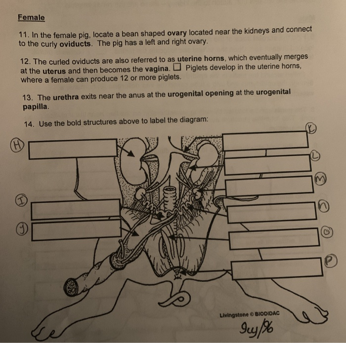
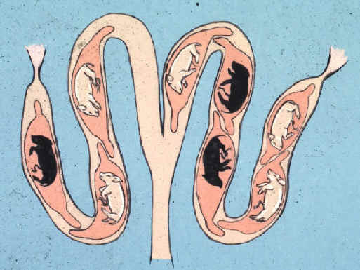
41 pig uterus diagram
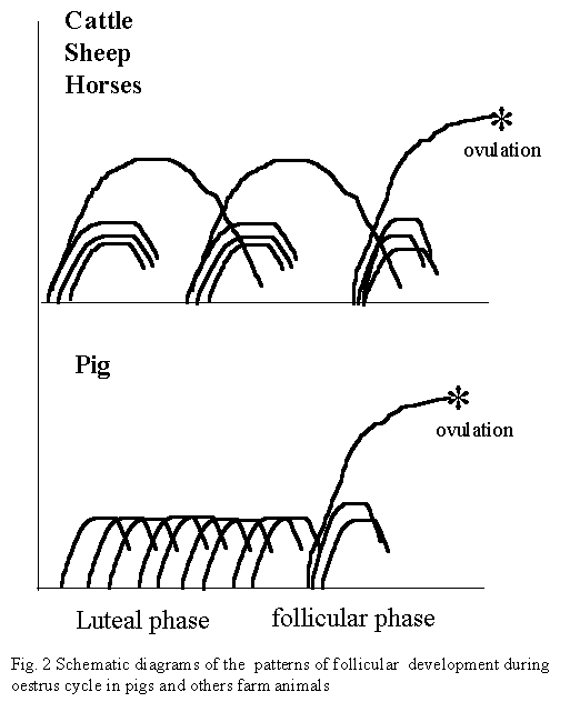
05.07.2021 · The horse of mare uterus is cylindrical and blunt at their cranial ends. Body of uterus located at abdominal cavity (partly in the pelvic cavity) and cylindrical, flattened dorsoventrally. The cervix is short, and the cervical canal is straight and simple. The endometrium is devoid of any cotyledon in the horse. The va(g)zi-nya of a horse is straight, shorter, and less capacious. You will not ... Epitheliochorial - maternal epithelium of the uterus comes in contact with the chorion, considered as primitive (pigs, ... pig; Pregnancy period (days) 18 – 21 21 – 23 110 – 118 Placenta type: Discoidal, decidual hemoendothelial choroidea Discoidal, decidual hemoendothelial choroidea Epitheliochorial Litter size 6 – 12 6 – 15 11 – 16 Birth weight (g) 0.5 – 1.5 3 – 5 900 ...
Welcome to the Whitman College Biology Department's Virtual Pig Dissection (VPD)! This site is designed as a supplement to laboratory dissections exploring introductory mammalian anatomy and physiology — it is basic and many details have been omitted for clarity. We hope that it is suitable for AP Biology students or for students of ...

Pig uterus diagram
Formerly guinea pig (Cavia sp.) (Fig. 19.1) were used for dissection in most of the undergraduate and postgraduate colleges in Indian Universities. Of late, due to prevailing high cost, guinea pig is being replaced by rat. Four species of rats are common in India, of which three are wild. Rattus rattus are the large-sized ones. R. rattus and R. bengalensis are commonly found in godowns ... Fetal Pig Dissection Lab Introduction: In this lab you will be examining many characteristics of an unborn mammal--the fetal pig. Dissection will help you to get a 3-dimensional picture of how all the systems fit together in an entire organism. You've seen separate diagrams of many of the major systems. Now you'll get to see how they are all arranged spatially. You'll also get a better idea of ... Fetal Pig Dissection Resources. To study the pig in more detail, go to this Virtual Pig Dissection. It covers all the body systems and includes quizzes to test your knowledge! Label the Anatomy of a Fetal Pig. Print out these PDFs and fill in the labels to test your knowledge of fetal pig anatomy. Internal anatomy: label the middle section
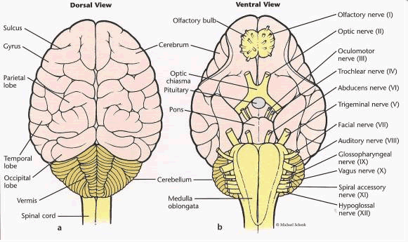
Pig uterus diagram. Diagram of the Genital Ducts at -the commencement of the 3rd month of foetal life. Lateral view. Fig. 87. Diagram of the Müllerian Ducts at the commencement of the 3rd month. Ventral view. Fig. 88. Evolution of the Human Form of Uterua. Fig. 89. Showing the manner in which the Mulleriau Ducts fuse to form the Uterus and Vagina. Fig. 90. A section of the Prostate showing the Hemnants of the ... Diagram of the spinal cord showing segments. The spinal cord is the main pathway for information connecting the brain and peripheral nervous system. Much shorter than its protecting spinal column, the human spinal cord originates in the brainstem, passes through the foramen magnum, and continues through to the conus medullaris near the second lumbar vertebra before terminating in a fibrous ... The fetal pig that you will dissect has been injected with a colored latex (rubber) compound. The arteries have been filled with red latex and the veins with blue. An incision was made on the side of the neck to enable the injections. The incision can be seen in the first photograph below. Several different pig dissections were used to obtain the photographs below. As a result, a structure ... 14.05.2021 · Rabbit internal anatomy diagram Now, I will show you some internal organ anatomy from rabbits with a diagram. Here, you will find the essential and most special anatomical features of the internal organs. Let’s start to know from the digestive organs of rabbit anatomy. Digestive organs of a rabbit. You will not find the muzzle in a rabbit ...
Fetal Pig Dissection Resources. To study the pig in more detail, go to this Virtual Pig Dissection. It covers all the body systems and includes quizzes to test your knowledge! Label the Anatomy of a Fetal Pig. Print out these PDFs and fill in the labels to test your knowledge of fetal pig anatomy. Internal anatomy: label the middle section Fetal Pig Dissection Lab Introduction: In this lab you will be examining many characteristics of an unborn mammal--the fetal pig. Dissection will help you to get a 3-dimensional picture of how all the systems fit together in an entire organism. You've seen separate diagrams of many of the major systems. Now you'll get to see how they are all arranged spatially. You'll also get a better idea of ... Formerly guinea pig (Cavia sp.) (Fig. 19.1) were used for dissection in most of the undergraduate and postgraduate colleges in Indian Universities. Of late, due to prevailing high cost, guinea pig is being replaced by rat. Four species of rats are common in India, of which three are wild. Rattus rattus are the large-sized ones. R. rattus and R. bengalensis are commonly found in godowns ...
Pig Blastocyst Uterine Interactions Sciencedirect

Reproductive System The Pig Site

Artificial Insemination In Swine How Do You Know When A Pig Is In Heat
The Reproduction In Pig
Human

Schematic Representation Of The Bicornuate Guinea Pig Uterus With Fetal Download Scientific Diagram
Solved 10 Why Does A Vasectomy Not Affect Urine Flow Be Chegg Com

Visceral Topography Of The Pregnant Sow 1 Gross Gut 2 Wide Download Scientific Diagram
2

Pig Blastocyst Uterine Interactions Sciencedirect

Artificial Insemination In Swine How Do You Know When A Pig Is In Heat
Pig Anatomy And Terminology Mini Pig Info

Ijms Free Full Text Integrating Lcm Based Spatio Temporal Transcriptomics Uncovers Conceptus And Endometrial Luminal Epithelium Communication That Coordinates The Conceptus Attachment In Pigs Html
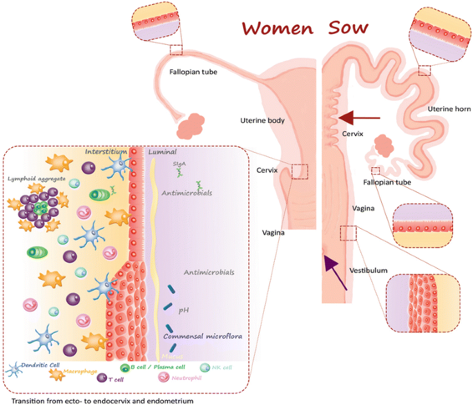
A Review Of The Human Vs Porcine Female Genital Tract And Associated Immune System In The Perspective Of Using Minipigs As A Model Of Human Genital Chlamydia Infection Veterinary Research
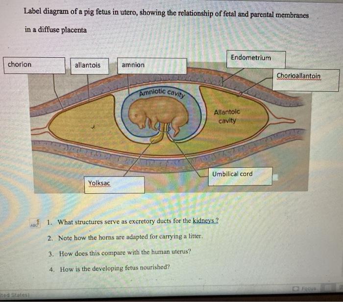
Solved Label Diagram Of A Pig Fetus In Utero Showing The Chegg Com
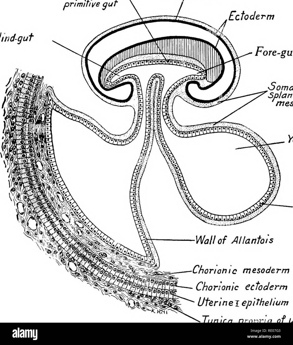
A Laboratory Manual And Text Book Of Embryology Embryology Lumbilicaj Vein L Vitellim Fein A Entoderm Of Gut Splanchnic Mesoderm Entoderm Of Primitive Gut 3 Amnio Ectoderm Hind Gut Fore Gut Somatic And Splanchnic Mesoderm

Anatomy Of The Sow S Uterus And Sperm Deposition Sites During Ai The Download Scientific Diagram
Sow Anatomy

Fetal Pig Reproductive System Female Flashcards Quizlet
Book Contributions To Embryology Carnegie Institution No 64 Embryology
2

Fetal Pig Dissection Work In Groups Of Ppt Video Online Download
2
Reproductive Physiology And Anatomy Of The Sow

Chordate Anatomy Chordata Anatomy Comparative Oviduc T Quot Uterus Quot Vagina D Fig 269 Four Types Of Uteri Occurring In Different Groups Of Mammals A Duplex The Type Found In Rodents B Bipartite

Advances In Artificial Insemination In Nulliparous Sows Articles Pig333 Pig To Pork Community
2

Ppt Sow Reproductive Tract Powerpoint Presentation Free Download Id 3104223

Variety Meat The Hidden Pork Value National Hog Farmer
Fetal Pig Dissection

The Decellularization Of The Pig Uterus Representative Pictures Download Scientific Diagram
1

Pig Anatomy Pt 4 Reproductive Anatomy Diagram Quizlet

The Anatomy Of The Domestic Animals Veterinary Anatomy The Stonlich 415 The Genital Fold Is Enlarged So As To Inclose The Uterus And A Small Part Of The Vagina It Forms
Artificial Insemination In Swine Breeding The Female Mu Extension

Uterus Bicornis An Overview Sciencedirect Topics

Parturition Farrowing The Pig Site
2

Cow Pig Uterus Diagram Diagram Quizlet
Urogenital System Mannix S Science Nerdery
2


Comments
Post a Comment