41 sacral plexus diagram
Diagram. Nerve to obturator internus and superior gemellus. Posterior femoral cutaneous nerve. From the case: Sacral plexus (diagram) Diagram. Posterior femoral cutaneous nerve. Perforating cutaneous nerve. From the case: Sacral plexus (diagram) Diagram. Sacral Bone Pain – Causes, Treatment, and Anatomy of Sacrum. Sacral bone pain is perceived in the lower part of the back and saddle area. Commonly, the underlying reason for pain in this region is injury or trauma to the joint present between the hip and vertebral column. Sometimes this pain can spread to other parts of the body and lead to ...
About Press Copyright Contact us Creators Advertise Developers Terms Privacy Policy & Safety How YouTube works Test new features Press Copyright Contact us Creators ...

Sacral plexus diagram
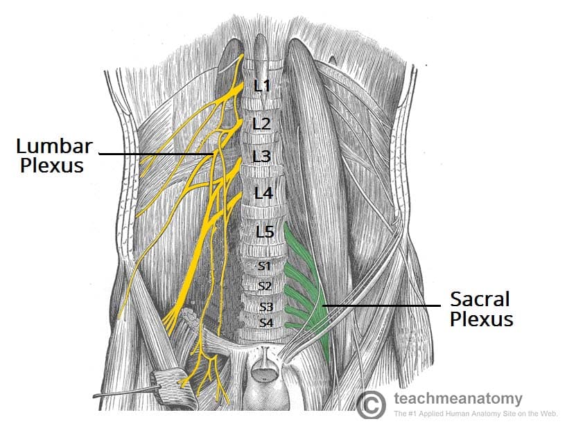
The sacral plexus comes from the lower lumbar nerves L4 and L5 and the sacral nerves S1 to S4. The most significant systemic nerve to come from this plexus is the sciatic nerve, which is a combination of the tibial nerve and the fibular nerve. Lumbar Plexus Diagram. ... network of nerves derived from the T12 through L4 spinal nerves. Both the lumbar and sacral plexus combine to form the lumbosacral plexus. There are six main branches of ... Fig. 11.1 Nerves of the lower extremity Fig. 11.2 Schematic diagram of the lumbar plexus Fig. 11.3 Lumbar and sacral plexuses within the skeleton Fig. 11.4 Major branches of the lumbar plexus The nerves of the lumbar plexus are responsible for supplying motor innervation to the transversus abdominis and internal oblique muscles, the cremaster muscle…
Sacral plexus diagram. The sacral plexus is a network of nerve fibres that supplies the skin and muscles of the pelvis and lower limb. It is located on the surface of the posterior pelvic wall, anterior to the piriformis muscle. The plexus is formed by the anterior rami (divisions) of the sacral spinal nerves S1, S2, S3 and S4. The sacral plexus is formed by the anterior rami of S1 to S4 as well as the lumbosacral trunk (anterior ramus of L4 & L5). The lumbosacral trunk courses ... Nerves of the sacral plexus: diagram This item is available to registered subscribers only Sign up now to obtain ten tokens to view any ten Vetlexicon articles, images, sounds or videos, or Login The anterior divisions of the lumbar, sacral, and coccygeal nerves form the lumbosacral plexus, the first lumbar nerve being frequently joined by a branch from the twelfth thoracic. For descriptive purposes this plexus is usually divided into three parts—the lumbar, sacral, and pudendal plexuses. The Lumbar Nerves (Nn.
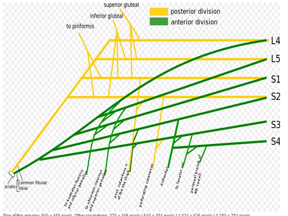
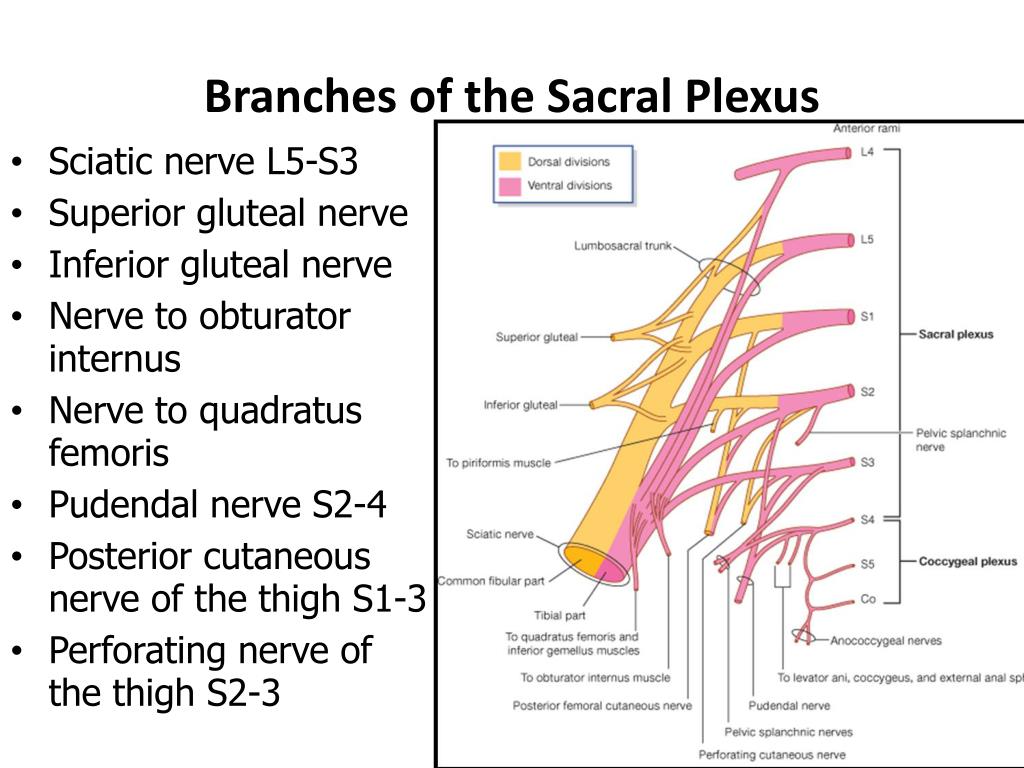
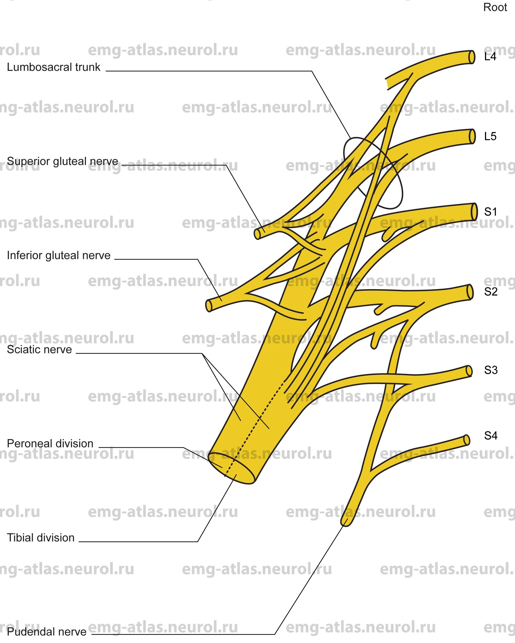
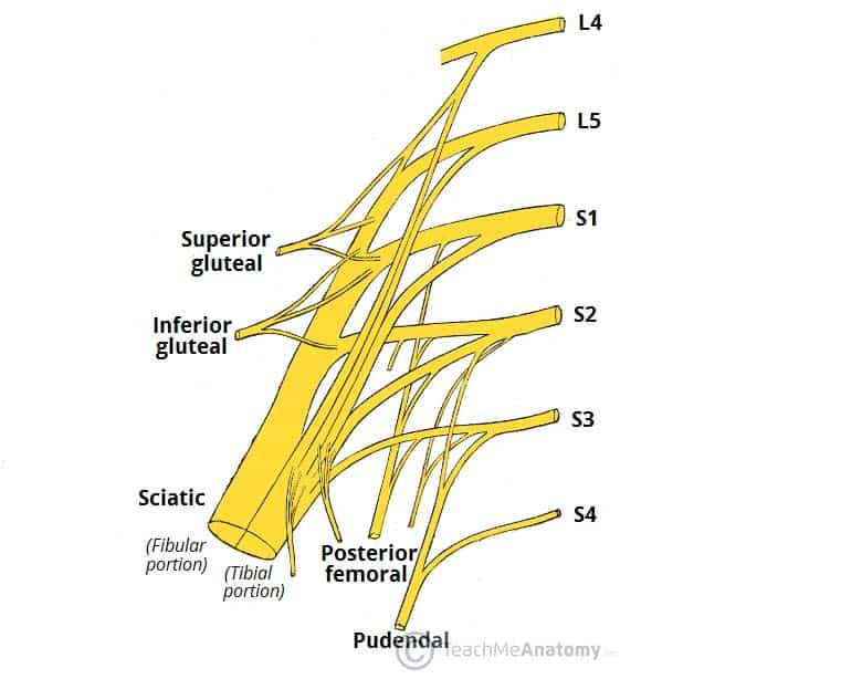
SACRAL NERVE PLEXUS ANATOMY Just inferior to the lumbar plexus lies the sacral plexus.The anterior rami of spinal nerves lumbar 4 and 5 as well as sacral 1 through 4 create the sacral plexus. The pelvis, the lower back, the perineum, the sections of the foot (dorsal and plantar) and sections of the thigh and leg ( posterior surface) all are innervated by the nerves that branch off the sacral ... The sacral plexus lies caudal to the lumbar plexus (stems from L4 to S4) and is often referred together withthe lumbar plexus as the lumbosacral plexus. The branches innervate buttocks, pelvis, perineum and lower limb (except for anterior and medial thigh). Sciatic nerve, the largest nerve of the sacral plexus is actually two nerves wrapped in ... The coccygeal plexus originates from the S4, S5, and Co1 spinal nerves. It is interconnected with the lower part of the sacral plexus. The only nerve in this plexus is the anococcygeal nerve, which serves sensory innervation of the skin in the coccygeal region. Sacral plexus diagram: Diagram of the sacral plexus showing the various anterior and ... Figure 13.11a The sacral plexus. Superior gluteal Lumbosacral trunk Inferior gluteal Common fibular Tibial Posterior femoral cutaneous Pudendal Sciatic (a) Ventral rami and major branches of the sacral plexus L 4 L 5 S 1 S 2 S 3 S 4 S 5 Co 1 Ventral rami Ventral rami:
Schematic diagram of the lumbar plexus. Lumbosacral plexus and sciatic nerve: diagrams. Case contributed by Mr Plan of sacral and pudendal plexuses. Author: Henry Vandyke. The lumbar plexus is a network of nerve fibres that supplies the skin and musculature of the lower limb. It is located in the lumbar region, within.The lumbar plexus is a ... They are typically named after the regions of the body they innervate also, such as the cervical, brachial, lumbar, sacral, celiac and coccygeal plexus. Auerbach's plexus is located in the GI tract. The sacral plexus is formed by anterior rami of L4 to S4 and its branches innervate the pelvis, perineum and lower limb.. Gross anatomy. The sacral plexus forms on the anterior belly of the piriformis muscle and is formed by the lumbosacral trunk (L4-5) of the lumbar plexus, which enters the pelvis coursing medially to psoas major and unites with the ventral rami of the S1 to S4 nerve roots ... The sacral plexus (plexus sacralis) is a nerve plexus that provides motor and sensory nerves for the posterior thigh, most of the lower leg, the entire foot, and part of the pelvis (see the following image). It is part of the larger lumbosacral plexus. The sacral plexus is derived from the anterior rami of spinal nerves L4, L5, S1, S2, S3, and S4.
Solved Drag And Drop 1 Sacral Spinal Nerves 2 Coccygeal Chegg Com
Sacral Plexus. Create healthcare diagrams like this example called Sacral Plexus in minutes with SmartDraw. SmartDraw includes 1000s of professional healthcare and anatomy chart templates that you can modify and make your own. 53/75 EXAMPLES. EDIT THIS EXAMPLE. CLICK TO EDIT THIS EXAMPLE.
Lumbar And Sacral Plexuses Ppt Video Online Download
Diagram of the sacral plexus, stressing the importance of the piriformis muscle. If the PM gets tense the sacral plexus doesn't have soft support anymore and symptoms of irritation of the sacral plexus may develop. The patient will exhibit a great variety of the clinical signs and symptoms because the sacral plexus is the origin of the majority ...
Lumbar Plexus Anatomy Branches And Innervation Kenhub
In human anatomy, the sacral plexus is a nerve plexus which provides motor and sensory nerves for the posterior thigh, most of the lower leg and foot, and part of the pelvis.It is part of the lumbosacral plexus and emerges from the lumbar vertebrae and sacral vertebrae (L4-S4). A sacral plexopathy is a disorder affecting the nerves of the sacral plexus, usually caused by trauma, nerve ...

Sacral Plexus Diagram Radiology Case Radiopaedia Org
Diagram - The sacral plexus, with the six spinal nerves on the left and the five peripheral nerves on the right (and the bottom). It is important to note that although in this diagram the sciatic nerve is shown to have two different portions with different roots, they make up the same nerve.

Sacral And Coccygeal Plexuses
Which plexus arises from the structure labeled B in the diagram? a) cervical plexus b) brachial plexus c) thoracic nerves d) lumbar plexus e) sacral plexus. a) A. In this diagram, respiratory distress can occur if damage occurs to which group of spinal nerves? a) A b) C c) E d) F e) H. a) E. In this diagram which structure is the conus medullaris?

Nerve Plexus Wikipedia
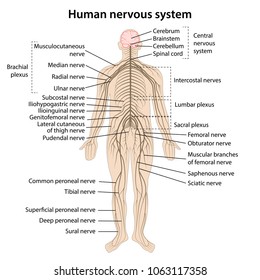
The sacral plexus supplies nerves to the posterior leg. Cranial Nerves. The nerves attached to the brain are the cranial nerves, which are primarily responsible for the sensory and motor functions of the head and neck (one of these nerves targets organs in the thoracic and abdominal cavities as part of the parasympathetic nervous system). There ...

Sacral Plexus Diagram 3
The lumbosacral plexus is formed by the anterior rami of the nerves (spinal segments T12-S4) to supply the lower limbs. The lumbosacral plexus can be divided into the lumbar plexus, which innervates the ventral upper half, and the sacral plexus, which mainly innervates the dorsal side. Image: The lumbar plexus and its branches.

The Lumbar And Sacral Plexuses Sciencedirect
The sacral plexus is a network of nerves emerging from the lower part of the spine. These nerves provide motor control to and receive sensory information from most of the pelvis and leg. A plexus is a web of nerves that share roots, branches, and functions. There are several plexi (plural of plexus) throughout the body, and the sacral plexus ...

Accessphysiotherapy Lumbar And Sacral Plexus With Clinical Cases
Another shortcut, hope it's helpful- God Bless

Accessphysiotherapy Lumbar And Sacral Plexus With Clinical Cases
Fig 6: Diagram showing the anatomical relationship between the piriformis muscle and the sacral plexus Finally, mainly small calibre (approx.1-2 mm) vegetative fibres, originating from S2, S3 and S4, interconnect to form a weblike network, located deep in the parametrium, to the side of the rectum and below the ureter: the inferior hypogastric ...

The Sacral Plexus Reproduced With Permission From Isaacs Re Fessler Download Scientific Diagram
Here, again I will provide the lumbosacral plexus nerves anatomy labeled diagram of an ox. I tried to show you the significant branches of the lumbosacral plexus of an ox. You will get the updated labeled diagram of the lumbar and sacral plexus of an ox, sheep, and goat on the social media of anatomylearner.com.

Plexus Products Study Solutions Anatomy Study
Mar 19, 2015 · Sacral plexus. A network of intersecting nerves is referred to as a nerve plexus. Nerves that serve the same part of the body merge into one large nerve or group of nerves via a plexus. The sacral ...

Lumbar And Sacral Plexus Flashcards Quizlet
1. Lumbosacral plexus, 2. Coccygeal plexus, and 3. Pelvic splanchnic nerves. Lumbosacral Plexus Formation. The lumbosacral plexus is formed by the lumbosacral trunk and the ventral rami of the first to third sacral nerves, and part of the fourth sacral nerve.

Sacral Plexus Anatomy Tutorial Youtube
Anatomy of the sacral plexus. Check out the 3D app at http://AnatomyLearning.com. More videos available on http://AnatomyZone.com.
Sacral Plexus Images Stock Photos Vectors Shutterstock
The brachial plexus is a network of nerves that gives rise to all the motor and sensory nerves of the upper extremity his plexus arises from the anterior rami of spinal nerves C5-T1 that undergo several mergers and splits into trunks and divisions, until they finally give rise to their terminal branchesThese terminal branches are responsible for motor and sensory innervation of the upper ...
Sacral Plexus Anatomy Branches And Mnemonics Kenhub
Sacral Plexus (Blank Diagram)

Lumbar And Sacral Plexus
Oct 28, 2021 · The sacral plexus is a nerve network comprised of the lumbosacral trunk and sacral spinal nerves. The lumbosacral trunk is formed by the lumbar spinal nerves L4 and L5. The trunk then descends into the pelvis to meet the roots of sacral spinal nerves S1 - S4, as they emerge from the spinal cord. Note that S4 root only partially contributes to the formation of the sacral plexus.

Lower Extremity Peripheral Nerve Blocks Lumbar Sacral Plexus Anatomy
Fig. 11.1 Nerves of the lower extremity Fig. 11.2 Schematic diagram of the lumbar plexus Fig. 11.3 Lumbar and sacral plexuses within the skeleton Fig. 11.4 Major branches of the lumbar plexus The nerves of the lumbar plexus are responsible for supplying motor innervation to the transversus abdominis and internal oblique muscles, the cremaster muscle…

Station 6 Lumbar And Sacral Plexuses Posterior Diagram Quizlet
Lumbar Plexus Diagram. ... network of nerves derived from the T12 through L4 spinal nerves. Both the lumbar and sacral plexus combine to form the lumbosacral plexus. There are six main branches of ...
1
The sacral plexus comes from the lower lumbar nerves L4 and L5 and the sacral nerves S1 to S4. The most significant systemic nerve to come from this plexus is the sciatic nerve, which is a combination of the tibial nerve and the fibular nerve.

Sacral Plexus Wikipedia

Schematic Drawing Of Lumbar And Sacral Plexus And The Main Pelvic Download Scientific Diagram
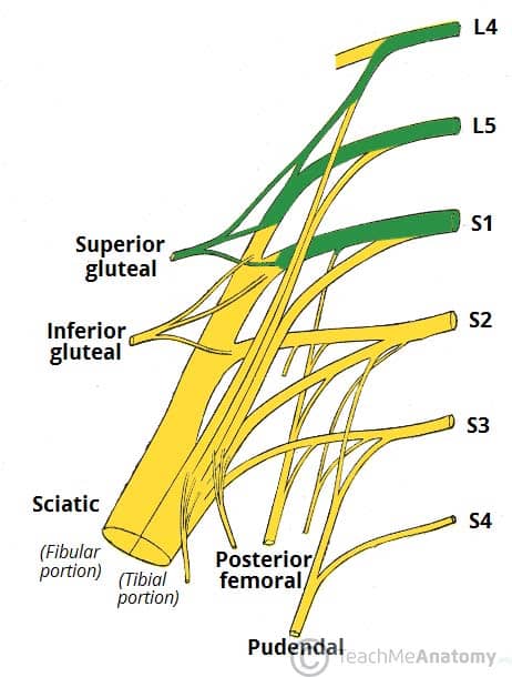
The Sacral Plexus Spinal Nerves Branches Teachmeanatomy

Sacral Plexus Diagram Quizlet

Distribution Of Spinal Nerves Boundless Anatomy And Physiology

12 7d Sacral And Coccygeal Plexuses Medicine Libretexts

Sacral Plexus Iliohypogastric Nerve Lumbar Plexus Ilioinguinal Nerve Sacral Plexus Text Hand Png Pngegg
Ppt Lumbosacral Plexus Powerpoint Presentation Free Download Id 2734304

File Sacral Plexus Schematic Svg Wikimedia Commons

Sacral Plexus And Derivative Nerves Muscles And Lesions Of The Lower Extremity Youtube

Diagram Of The Sacral Plexus General Practice Notebook
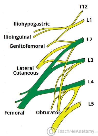
The Lumbar Plexus Spinal Nerves Branches Teachmeanatomy

Sacral Plexus Diagram Quizlet

How To Draw Sacral Plexus Lower Limb Doctor Z Youtube
Online Atlas Of Electromyography

Sacral Plexus Diagram Quizlet

Relationships Between Sacral Plexus And Superior Gluteal Artery Download Scientific Diagram
The Sacral Plexus Spinal Nerves Branches Teachmeanatomy
1

Sacral Plexus Roots Branches Innervation Youtube
The Sacral Plexus Spinal Nerves Branches Teachmeanatomy
Comments
Post a Comment