42 sponge diagram labeled
Oct 14, 2019 · Build the front assembly by placing the front slats (A) against the assembly frame. Glue and nail the corner cleats (C) with their outside edges flush with the slat ends (Project Diagram, Drawing 1) so that the bottom ends of the braces overhang the bottom edge of the sides by 3/4 inch (Photo 2).Remove the assembly and build the back using the same parts. Reliable, wide range, and highly sensitive joint movement monitoring is essential for training activities, human behavior analysis, and human-machine interfaces. Yet, most current motion sensors work on the nano/microcracks induced by the tensile deformation on the convex surface of joints during joint movements, which cannot satisfy requirements of ultrawide detectable angle range, high angle ...
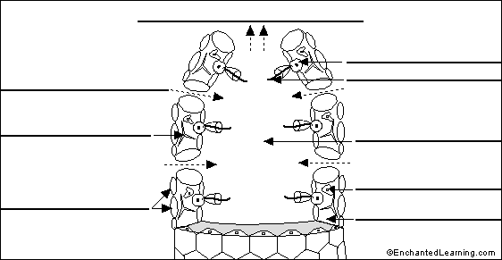
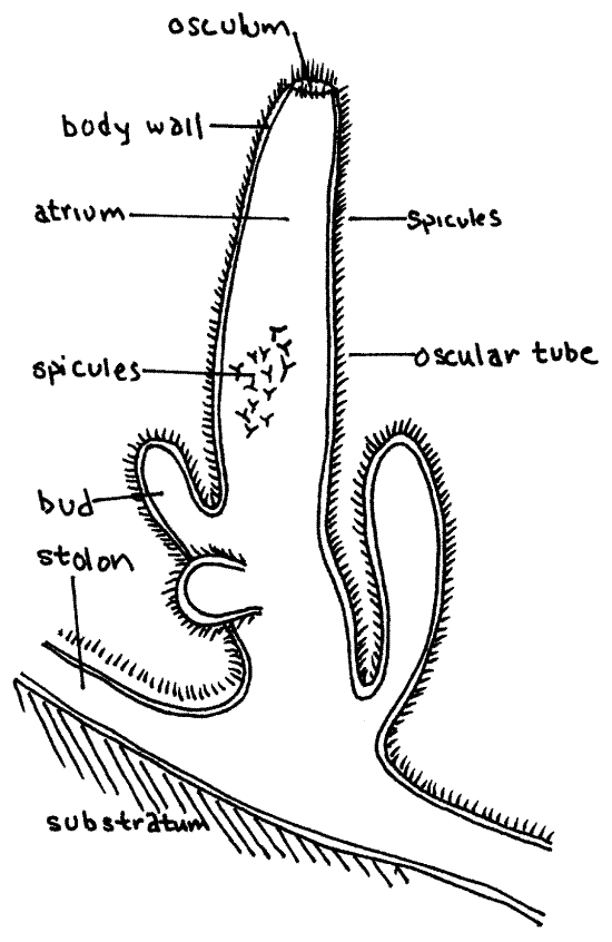
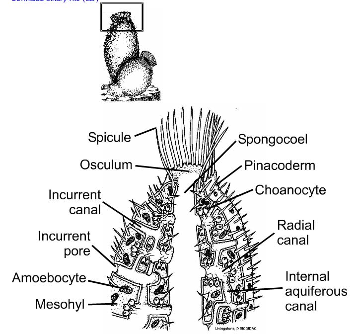
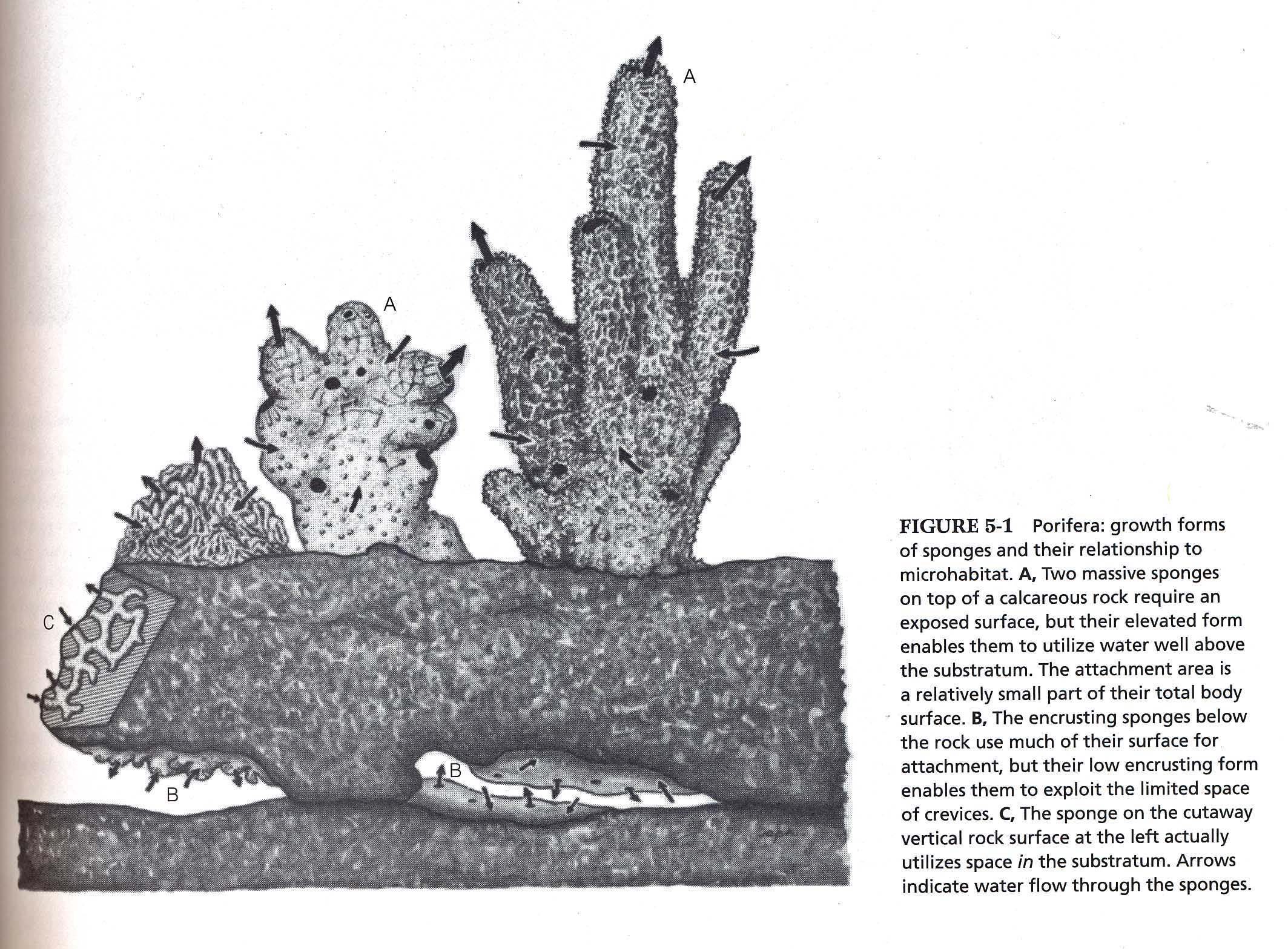
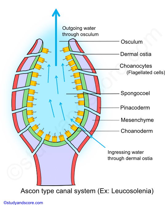
The following diagrams come from Invertebrate Zoology, by Rupert and Barnes. The photos are my own. Here is a close view of an asconoid sponge from our aquaria in the lab - similar if not identical to the one in Fig 5-3 B above:
Sponge diagram labeled
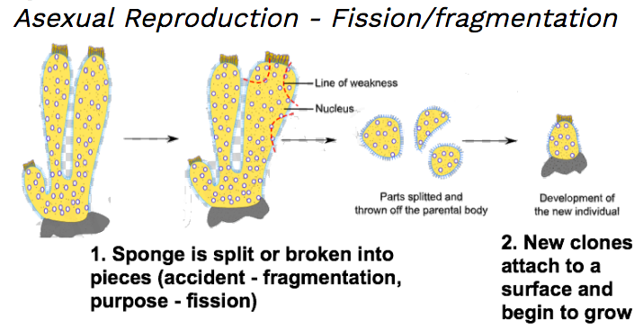
5. Variety in form (see diagrams in web article) asconoid; syconoid; leuconoid (most common) 6. Cool things sponges can do: many can move to new locations by dissassembly-reassembly (see: chicken liver sponges climbing up turtle grass) most sponges canbe disaggregated and cells can reaggregate into new sponge. 7. Ecological considerations Answer pages are only available to subscribers of EnchantedLearning.com . To subscribe to EnchantedLearning.com, click here (Fig. 9.3(b) is adapted from Phase Diagrams of Binary Nickel Alloys , P. Nash (Ed.), ASM International, Materials Park, OH, 1991.) Phase Diagrams: weight fractions of phases wt% Ni 20 1200 1300 T(°C) L (liquid) α L + α (solid) l i q u i d u s s o l i d u s 30 40 50 L + α Cu-Ni system TA A 35 Co 32 CL B TB D TD tie line 4 Cα 3 R S At TB ...
Sponge diagram labeled. The Trades menu can be accessed by opening the SkyBlock Menu, then by clicking the central Emerald on the menu, labeled "Trades". A new menu will open. Here, you can see the items which you can trade for. Slots filled with Grey Dye are currently locked, meaning that the player needs to unlock them, likely through a Collection unlock. The player ... 2. Draw, label, and color the “Sponge Morphology” diagram on the back of the paper (top half). 3. Define the following terms: a. Body wall – b. Ostia – c. Filter chambers – d. Atrium – e. Osculum – 4. Describe the path of water flow through a sponge. 5. Why is water flow important for sponges? 6. What are collar cells? Nov 06, 2020 · Stir the stain thoroughly to ensure color consistency. Using the 2” foam brush, apply a liberal coat of stain to the panels labeled 1 on “Diagram A.” Be sure to work the stain well into the embossed grain pattern. All corners and crevices of each work area should also be coated. A variation diagram is a plot showing how each oxide component in a rock varies with some other oxide component. Because SiO 2 usually shows the most variation in any given suite of rocks, most variation diagrams plot the other oxides against SiO 2 as shown in the diagram here, although any other oxide could be chosen for plotting on the x-axis.
Scypha, also known as crown sponge, is a small, marine sponge found attached by a sticky secretion to some submerged solid object like rocks, shells of molluscs and corals. It is found in shallow water up to a depth of 50 fathoms (1 fathom = 6 feet) where waves provide the animal with plenty of food and well oxygenated water. To help students learn the anatomy of the sponge, print out a black and white version of the color diagram below: Anatomy of the Sponge. _____ Testing and Assessment. Assess content comprehension about Porifera (Sponges) with the Mutiple Choice Test. Assess anatomical vocabulary comprehension of the Sponge Anatomy Labeling Page. The clitoris (/ ˈ k l ɪ t ər ɪ s / or / k l ɪ ˈ t ɔːr ɪ s / ()) is a female sex organ present in mammals, ostriches and a limited number of other animals.In humans, the visible portion – the glans – is at the front junction of the labia minora (inner lips), above the opening of the urethra.Unlike the penis, the male homologue (equivalent) to the clitoris, it usually does not ... A point on the diagram represents a composition that is specified in terms of mole fraction or weight fraction. The point, (0.3, 0.4, 0.3) is at the center of the small triangle in the diagram and is located by following the red diagonal 60° line at red 0.3 and the horizontal line at blue 0.4 or any combination of two of the coordinates (A, B, C).
Correctly label the following features of sponge anatomy. I pore osculum w H.O out nucleus sponge wall spicule arnoebocyte epidermal cell H.O in through pores collar cell (choanocyte) amoebocyte collar central cavity flagellum < Prey 5 of 18 18 Next > Question: Correctly label the following features of sponge anatomy. I pore osculum w H.O out ... Axial skeleton and pectoral girdle. 38 terms. Betsy_Gore TEACHER. Bone Cells. 20 terms. Betsy_Gore TEACHER. THIS SET IS OFTEN IN FOLDERS WITH... Sponges Review. 30 terms. Sponge flies, also known as spongilla-flies ... as shown in the diagram. Several sponge species are able to convert coral-derived DOM into sponge detritus, ... or even a completely separate kingdom of life, labeled Archaeata or Inferibionta. Since the 1990s archaeocyathids have been regarded as a distinctive group of sponges. = skin = aragonite Back to the water cycle diagram for students. Animals. The water cycle is critical to all life on Earth. View full size. In the grand scheme of the water cycle, animals may not play a very big role, but all animals, including you, participate in moving water around as part of the water cycle. ... to get a sponge to soak up the liquid before it ...
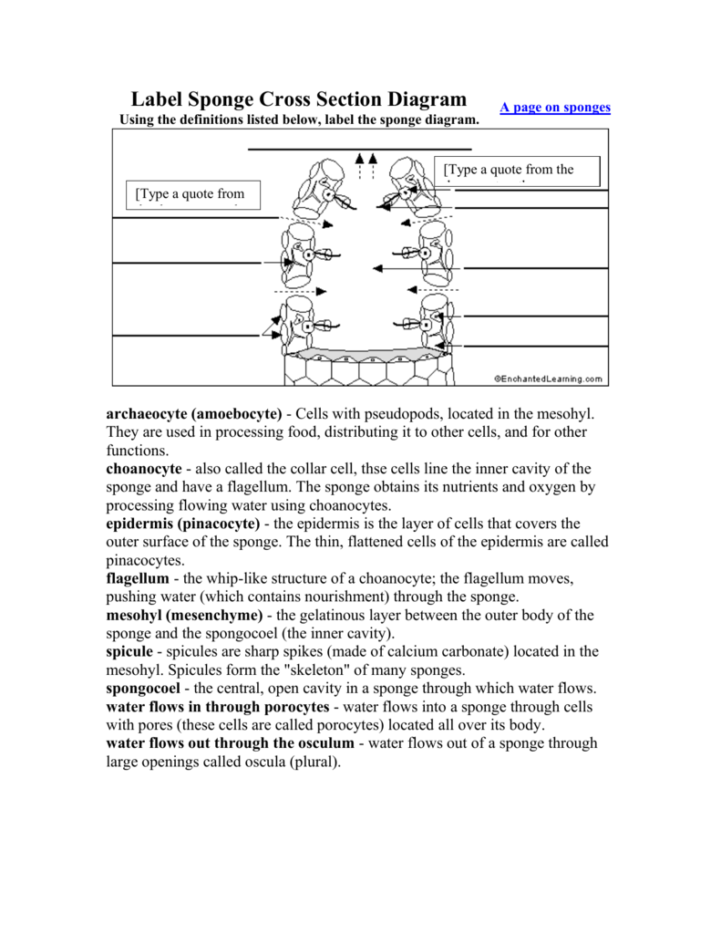
Label Sponge Cross Section Diagram Blank
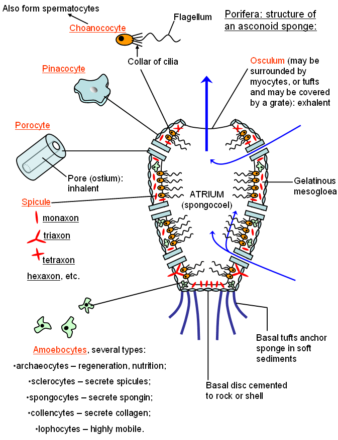
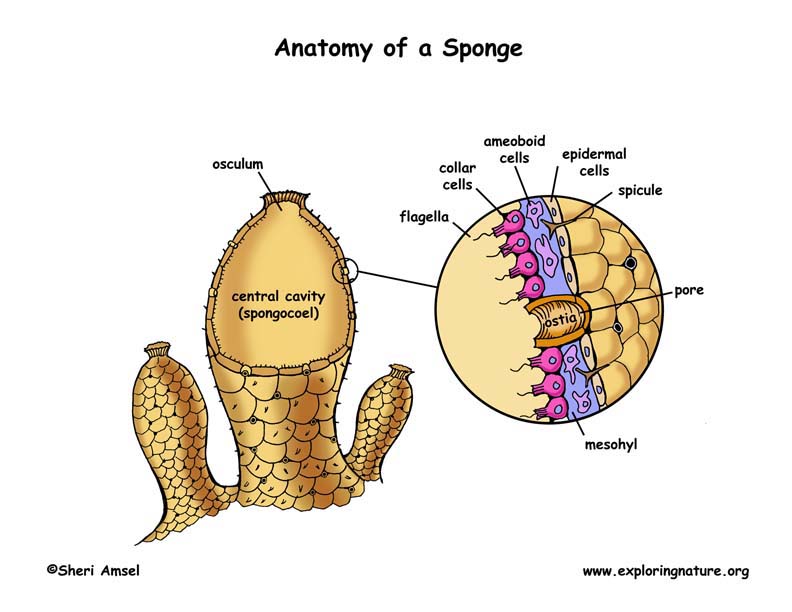
Label Sponge External Anatomy Diagram Using the definitions listed below, label the sponge and the flow of water through it. A page on sponges: epidermis - the layer of cells that covers the outer surface of the sponge. The thin, flattened cells of the epidermis are called pinacocytes.

Untitled Page
The primitive structure of a sponge consists of only two layers of cells separated by a non-living jelly like substance. The outer layer of the sponge is the epidermis which is made of flat cells called epithelial cells. Color all the epithelial cells (B) of the epidermis peach or pink. The inner layer consists of collar cells (A) whose ...

Sponge Structure And Function Advanced Read Biology Ck 12 Foundation
(Fig. 9.3(b) is adapted from Phase Diagrams of Binary Nickel Alloys , P. Nash (Ed.), ASM International, Materials Park, OH, 1991.) Phase Diagrams: weight fractions of phases wt% Ni 20 1200 1300 T(°C) L (liquid) α L + α (solid) l i q u i d u s s o l i d u s 30 40 50 L + α Cu-Ni system TA A 35 Co 32 CL B TB D TD tie line 4 Cα 3 R S At TB ...

Sponges And Cnidarians Concepts Of Biology
Answer pages are only available to subscribers of EnchantedLearning.com . To subscribe to EnchantedLearning.com, click here
Label Sponge External Anatomy Enchantedlearning Com
5. Variety in form (see diagrams in web article) asconoid; syconoid; leuconoid (most common) 6. Cool things sponges can do: many can move to new locations by dissassembly-reassembly (see: chicken liver sponges climbing up turtle grass) most sponges canbe disaggregated and cells can reaggregate into new sponge. 7. Ecological considerations

Life Cycle Animal Biology Spring 2010

Biology 11
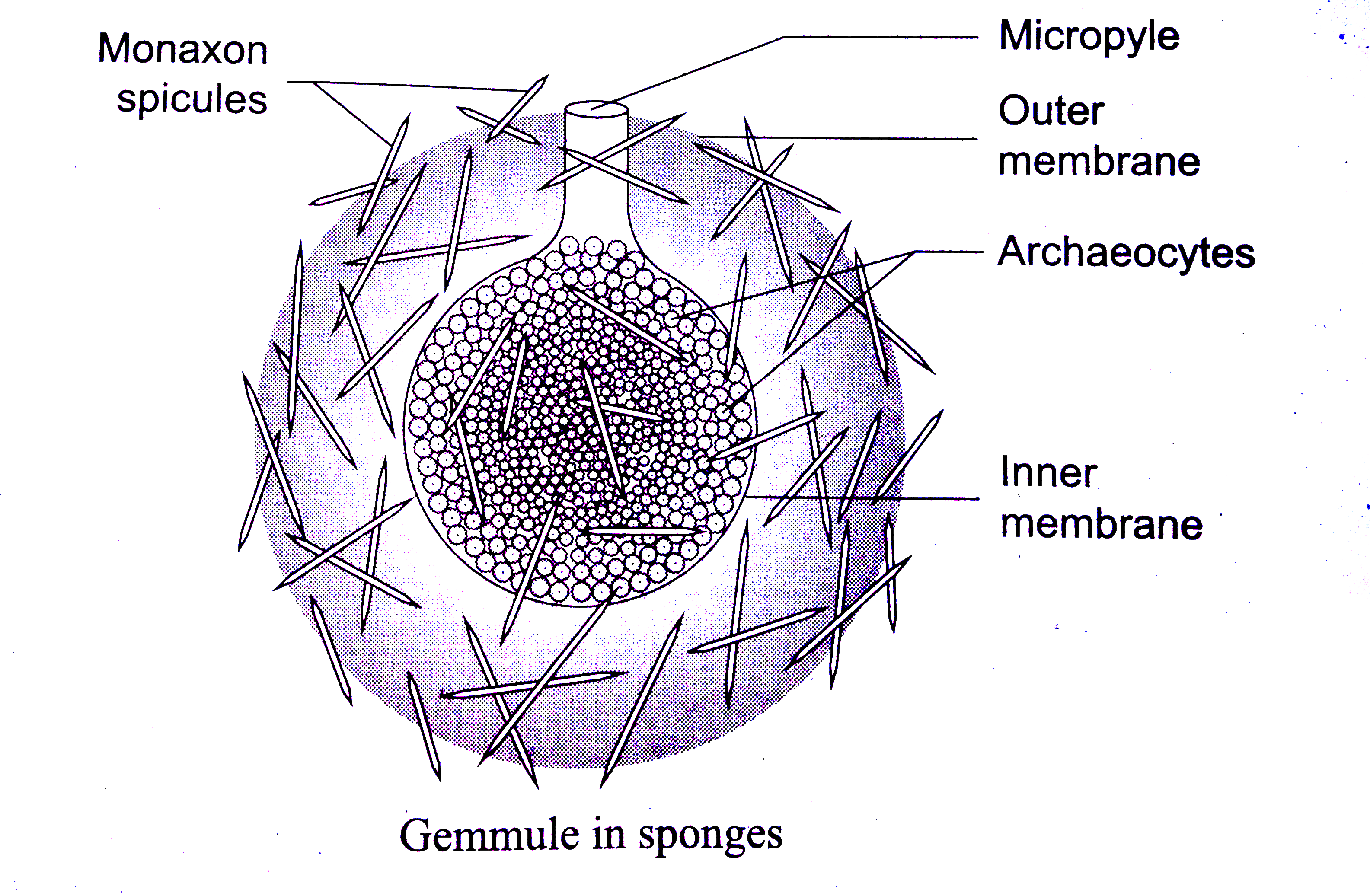
Draw And Label A Gemmule Of Sponge

Untitled Page

Sponge Wikipedia

Mr Joanides Wiki Pages Licensed For Non Commercial Use Only Porifera And Cnidarians 6 Biology Facts Anatomy Biology Notes

Use The Diagram Above To Answer The Following Questions 1 5 An Answer May Be Used More Than Once Or Not At All 1 Which Structure Would Be Course Hero
Sea Sponges Sea Sponges Diagram Unbelievable Facts

Types Of Locally Labeled Sea Sponges A Drawing Of A Vase Shaped Download Scientific Diagram
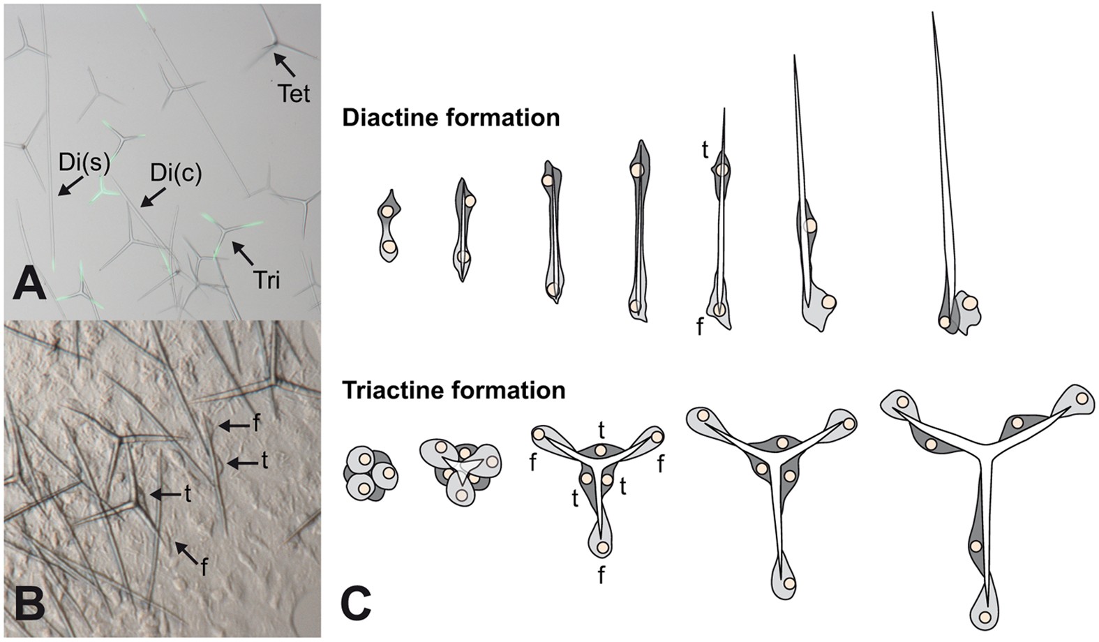
Spicule Formation In Calcareous Sponges Coordinated Expression Of Biomineralization Genes And Spicule Type Specific Genes Scientific Reports
Phylum Porifera

Sponge Gross Anatomy And Distinguishing Features Traits Unique To Download Scientific Diagram
Unit 5 1 Phylum Porifera The Biology Classroom
1

Sponge Diagram Labelled Synergy Middle School Science 08 09
How To Draw Sycon Sponge Labelled Diagram Kingdom Animalia Phylum Porifera Youtube
1

3 Diagrammatic Representation Of A Simple Asconoid Sponge Download Scientific Diagram
Label Sponge Cross Section Enchantedlearning Com

Systematic Biology

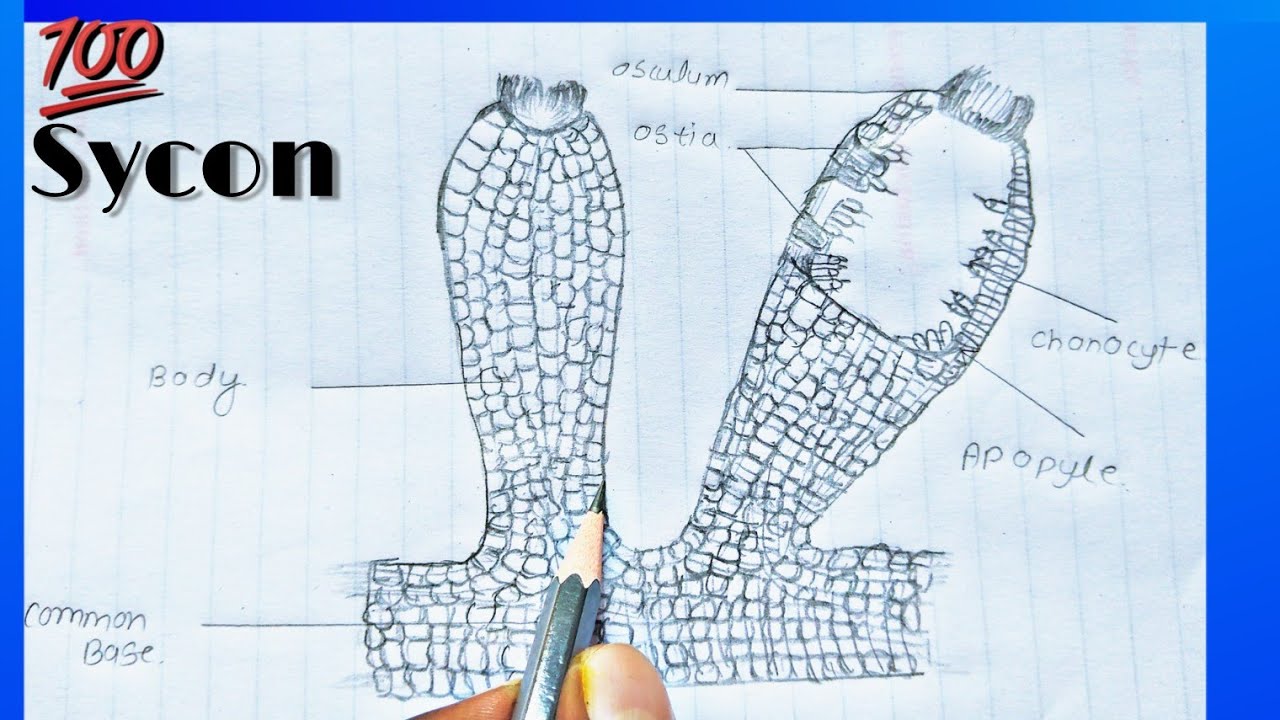
Scypha History Habitat And Nutrition With Diagram
Untitled 1
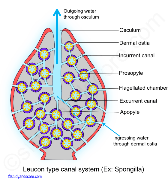
Phylum Porifera Canal System In Sponges Types Of Canal Systems In Sponges Study Score
Porifera
Phylum Porifera Sponges

Sponge Anatomy Diagram Quizlet

Marine Bio Ch 8 Sponge Label Diagram Quizlet
Overview Of Sponges

Sponge Labeling Diagram 1 Pdf Sponge Labeling Diagram Poriferans Pore Bearing Sponges Are Invertebrates Have No Backbone Or Notochord Once Course Hero
Sycon Sponge Clipart Etc
Bio385 Porifera
Sponge Diagrams And Photos
Phylum Porifera Canal System In Sponges Types Of Canal Systems In Sponges Study Score

Sponge Structure And Function Advanced Read Biology Ck 12 Foundation
Sponge

Adw Spongetypes Jpg

Sponge Diagrams Flashcards Quizlet


Comments
Post a Comment