40 cat blood vessel diagram
Start studying Cat Vessels. Learn vocabulary, terms, and more with flashcards, games, and other study tools. Circulatory System. Cat Upper Neck Arteries Veins Diagram Quizlet. Cat Veins Purposegames. Https Massasoit Instructure Com Courses 902777 Files 29763690 Download Wrap 1. Anatomy Lab Practicum 3 Cat Arteries And Veins Diagram Quizlet. Diagrams Cat Veins And Arteries Diagram. Untitled Document. Labeled Cat Veins Diagram.
lateral recumbency and to occlude the vessel. ii. Location 1. Typical site in cats. 2. Between the stifle and the femoral triangle. iii. Place pressure over the proximal vessel, just below the coxofemoral joint, using the lateral side of the hand. iv. Grasp the leg from the underside and start as far distally on the leg as possible. v.

Cat blood vessel diagram
VCD - Vessels. This is the first part of the cat where actually cutting will take place. Students will need to open the thoracic (chest) cavity to reveal the heart and its attached vessels. The best tool for this job is a scalpel and bone cutters to break the sternum. Once the heart is revealed, you can identify the vessels that attach to it. Navigate to the Cardiovascular System area in the following PAL 3.0 module: Human Cadaver, Anatomical Models, Histology, Cat, and Fetal Pig. MP3 Tutor Sessions Factors Regulating Blood Pressure. Interactive Physiology with Quizzes Cardiovascular System: Anatomy Review: Blood Vessel Structure and Function Cardiovascular System: Measuring Blood ... The lymphatic system also works with the cardiovascular system to return fluids that escape from the blood vessels back into the blood stream. The digestive system ( cat ) ( dog ) includes the mouth, teeth, salivary glands, esophagus, stomach, intestine, pancreas, liver and gall bladder.
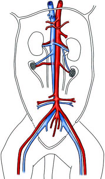
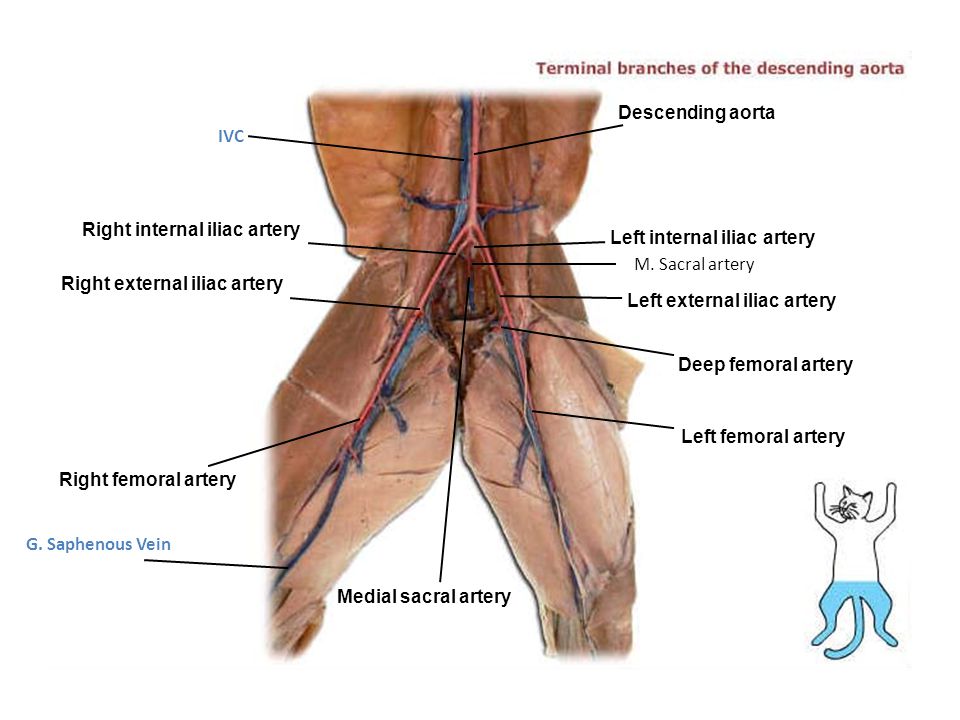
Cat blood vessel diagram. Renal arteries. External iliac arteries. Femoral vein. Femoral arteries. Internal iliac arteries. Common iliac veins. Abdominal aorta. Renal veins.80 pages Labeling Exercises for Blood Vessels Labeling Exercises for the Cat Blood Vessels and Sheep Heart Heart Diagram for Coloring Animation: Autorhythmic Cells Cardiac Action Potential Animation: Complete Review of the Heart Animation: Conducting System of the Heart Animation: The Cardiac Cycle Animation: The Cardiac Cycle Part II LEARNING OUTCOMES: ❍ Identify the major arteries and veins of the feline cardiovascular system. ❍ Compare the blood vessels of the cat with those of ... A thrombus is a blood clot that develops within the heart or a blood vessel. An embolus is a blood clot that arises in one area of the circulatory system and is transported in the bloodstream to a distant site, where it becomes lodged in a blood vessel. The most common form of this disease in cats is the development of a blood clot in the left ...
Cat Circulation. Click on a number below to see blood vessels in the area specified. 1. Carefully clear away any thymus tissue or fat obscuring the heart and the large vessels associated with the heart. Before identifying the blood vessels, try to locate the phrenic nerve (from the cervical plexus), which innervates the diaphragm (this may be quite difficult in your cat so do not spend an inordinate amount of time in this pursuit). This is an online quiz called Cat Arteries. There is a printable worksheet available for download here so you can take the quiz with pen and paper. From the quiz author. Cat Arteries Your Skills & Rank. Total Points. 0. Get started! Today's Rank--0. Today 's Points. One of us! Game Points. 26. Veins And Arteries Diagram Ap Cat Veins And Arteries Diagram Quizlet. Veins And Arteries Diagram 201 Structure And Function Of Blood Vessels Anatomy And Physiology. Veins And Arteries Diagram Coronary Artery Pass Graft Cabg How Its Performed Nhs. Veins And Arteries Diagram Box Plot Trends For Blood Flow Velocity In Arteries And Veins A
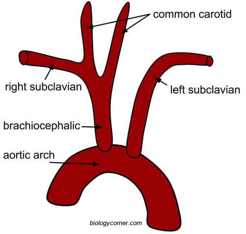
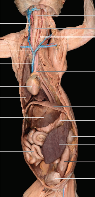
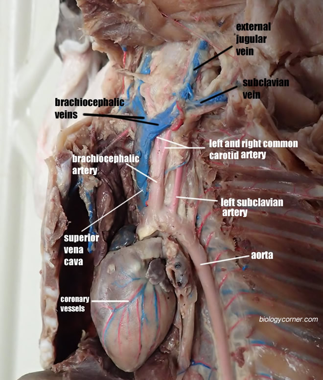
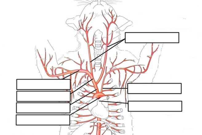
Investigation: Blood Vessels above the Diaphragm. Double-injected cats are usually used to identify blood vessels. Arteries are injected with red latex, and veins are injected with blue latex. ... Color code the diagram below (the aorta is the large vessel on the right) with red for artery and blue for vein. Cardiovascular System of the Cat. The pictures in this section are reprinted with permission by the copyright owner, Hill's Pet Nutrition, from the Atlas of Veterinary Clinical Anatomy. These illustrations should not be downloaded, printed or copied except for personal, non-commercial use. The cardiovascular system includes the heart and blood ... Cat Arteries And Veins 5 Diagram Quizlet. Solved Observe The Human Torso Model And Figures 63 7 63 9 A. Abdominal Cat Arteries. The Anatomical Record Anatomy Anatomy Vascular Abnormalities In. Ppt Inferior Vessels Of Cat Powerpoint Presentation Free. Lab Quiz 3b Cat Body Veins Arteries Diagram Quizlet. Vetcheck. Cat Arteries & Veins – Lab. This artery off the Aortic Arch is the Brachiocephalic, and will split to 3, what are they? This artery picked up, is the Celiac ...
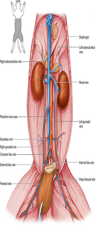
Cat Blood Vessels Of Abdominal Cavity Diagram Quizlet
Cats are known to have about 24 whiskers on their muzzle. Each whisker is deeply rooted in their body and reaches the very base where there are loads of blood vessels and nerves to draw from. It may shock you to learn that a single whisker of a cat is served by more than 200 nerve cells.
2
Identify the blood vessels indicated by the arrows on the dissected cats. Arteries (red) Veins (blue) Aorta. Brachiocephalic Artery. Common Carotid Artery. Descending Aorta. External Iliac Artery. Femoral Artery.

Cat Veins Quiz
Investigation: Blood Vessels above the Diaphragm. Double-injected cats are usually used to identify blood vessels. Arteries are injected with red latex, and veins are injected with blue latex. Blood vessels differ slightly in location from cat to cat. Carefully remove the fascia with blunt instruments to separate blood vessels from other ...

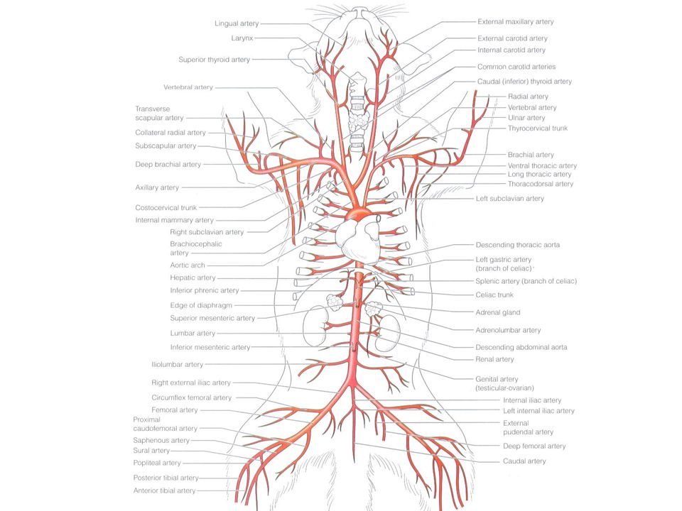
Key To The Arterial Anatomy Of The Cat Medical School Studying Medical Anatomy Arteries Anatomy
Cat Vessels Image Gallery. During this lab, students carefully teased the muscles and tissues away to reveal the arteries and veins. The goal was to identify each of the vessels on their lab guide and pass a lab test at the end of the activity where they are asked to locate the vessels.. After a Y incision is made, students carefully remove the pericardium and tissue surrounding the heart so ...

Major Veins And Arteries Of Cat Upper Thorax Quiz
5. Prepare a label for your cat with the names of your group members and the gender of your cat. 6. Follow the instructions for skinning the cat if you are dissecting skeletal muscles,or the instructions for opening the ventral body cavities if you are dissecting an organ system. FIGURE CP.1Directional terminology for the cat. Incision line

Arteries In The Human Body Anatomy Stock Illustration Download Image Now Istock
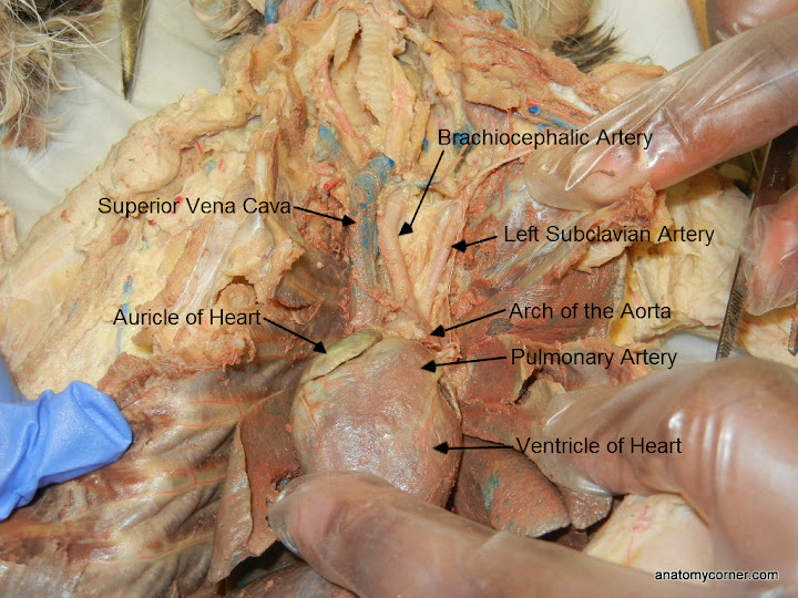
One of the first tasks of the cat dissection is to identify the vessels of the cat. We remove the pericardium from the heart and carefully tease away the connective tissue to reveal the arteries that leave the heart. If you follow the aortic arch, the first main vessel of the cat is the brachiocephalic artery.

Left Gastric Artery An Overview Sciencedirect Topics
Professor Fink reviews CAT Blood Vessels; incl. Aortic Arch; Brachiocephalic Artery; Celiac & Mesenteric Arteries; Inferior Vena Cava; Iliac Artery & Vein; B...

Arteries Concise Medical Knowledge
Blood Vessels Definition. The blood vessels are an intricate network of hollow tubular structures carrying blood throughout the body. They transport blood cells, nutrients and oxygen and carry away carbon dioxide and waste materials from the tissues and organs. The study of blood vessels is called angiology.

Quotes About Arteries 73 Quotes
Not all inclusive, check your study sheets for complete list. Learn with flashcards, games, and more — for free.
2
In the lungs, blood absorbs oxygen and gives up carbon dioxide. The blood then flows through the pulmonary veins into the left atrium. When the left ventricle ...

Jennifer Kersey E Portfolio Bio211 2011 04 10 Vet Medicine Anatomy Vet Tech School
This is an online quiz called Cat Arteries and Veins. There is a printable worksheet available for download here so you can take the quiz with pen and paper. Your Skills & Rank. Total Points. 0. Get started! Today's Rank--0. Today 's Points. One of us! Game Points. 43.
Cat Vessels Image Gallery
6 Biol 111 - Lab 9: Cat Circulation Name the vessel that is supplying blood to and from the vessel that is draining each of the following structures. Supplying Blood Structure Draining Blood Kidney Hind limb Pectoral muscles Head Front limb
Solved Observe The Human Torso Model And Figures 63 7 63 9 And Chegg Com
Looking at the major arteries and veins seen on Anatomy models and on some dissected (and plastinated) cats.
Introduction To Heart And Blood Vessel Disorders In Cats Cat Owners Merck Veterinary Manual
Diagram: Trick to remember the function of the left side of the heart is to pump oxygenated blood to the rest of the body - Blood that has "LEFT" to the lungs. Blood Flow of the Heart Review Let's now use the 2x2 table we made in the anatomy of the heart post, and this will give us another way to visualize the blood flow through the heart.

The Anatomical Record Anatomy Anatomy Posterior Cardinal Veixs Ix Ax Adult Cat 125 Ent For A Very Short Distance The Posterior Cardinals Converge And Empty Into This Both Renal Veins Instead
The lymphatic system also works with the cardiovascular system to return fluids that escape from the blood vessels back into the blood stream. The digestive system ( cat ) ( dog ) includes the mouth, teeth, salivary glands, esophagus, stomach, intestine, pancreas, liver and gall bladder.
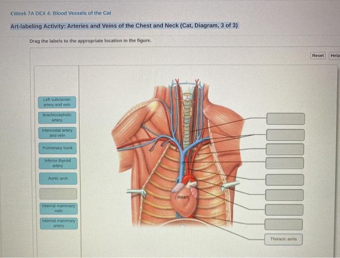
Solved Week 7 A Dex 4 Blood Vessels Of The Cat Chegg Com
Navigate to the Cardiovascular System area in the following PAL 3.0 module: Human Cadaver, Anatomical Models, Histology, Cat, and Fetal Pig. MP3 Tutor Sessions Factors Regulating Blood Pressure. Interactive Physiology with Quizzes Cardiovascular System: Anatomy Review: Blood Vessel Structure and Function Cardiovascular System: Measuring Blood ...
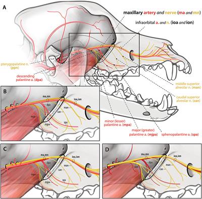
Frontiers Ligation Of The Maxillary Artery Prior To Caudal Maxillectomy In The Dog A Description Of The Technique Retrospective Evaluation Of Blood Loss And Cadaveric Evaluation Of Maxillary Artery Anatomy Veterinary
VCD - Vessels. This is the first part of the cat where actually cutting will take place. Students will need to open the thoracic (chest) cavity to reveal the heart and its attached vessels. The best tool for this job is a scalpel and bone cutters to break the sternum. Once the heart is revealed, you can identify the vessels that attach to it.
Understanding Heart Disease
2
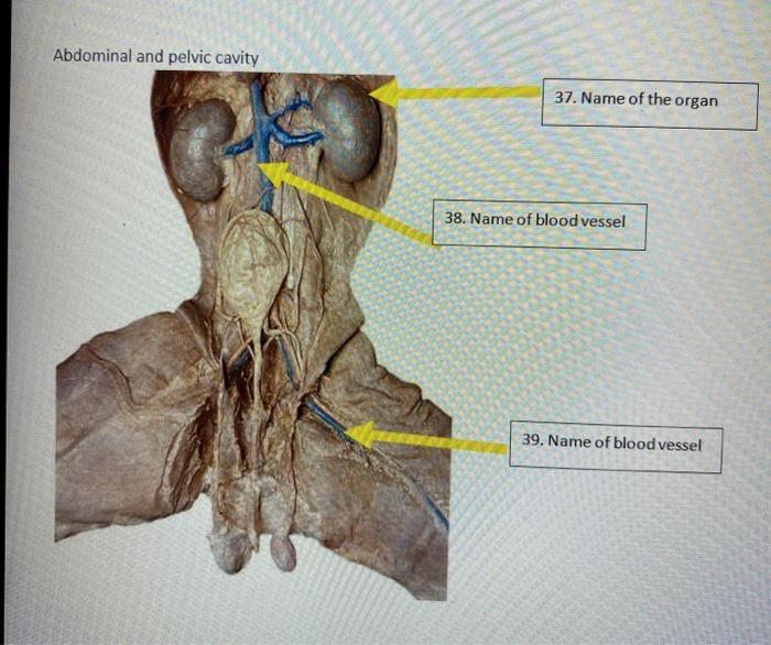
Solved Abdominal And Pelvic Cavity 37 Name Of The Organ 38 Chegg Com

Cat Femoral Artery Anatomy Limb Cat Angle Animals Anatomy Png Pngwing
Vcd Vessels
Dissection Of Blood Vessels Of Cat Flashcards Easy Notecards
Cat Dissection
1

Pin By Tara Chimenti On Vet Tech Vet Medicine Veterinary Technician Student Vet Tech Student

Cat Arteries Veins Diagram Diagram Quizlet

Cat Dissection Blood Vessels Flashcards Quizlet

An Update On Cerebrovascular Disease In Dogs And Cats Veterinary Clinics Small Animal Practice
1
Inferior Vessels Of Cat Ppt Video Online Download
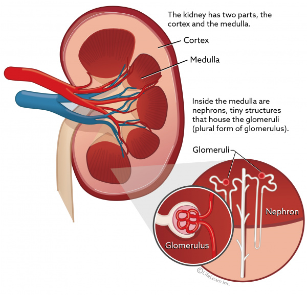
Glomerulonephritis In Cats Vca Animal Hospital
Cat Vessels Image Gallery
Cat Circulatory System Lab Guide
Cat Dissection

Anatomyforme Diagrams Of Feline Arterial And Venous Systems Vet Tech School Vet Medicine Veterinarians Medicine
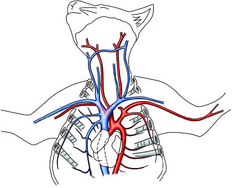
Cat Circulatory System Brachiocephalic Vein External Jugular Vein Common Carotid Artery Subclavian Artery And Vein Ppt Download

Upper Body Cat Blood Vessels Diagram Quizlet

Cat Arteries Complete Overview Diagram Quizlet
1


Comments
Post a Comment