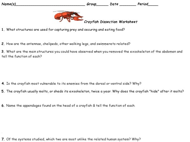
40 crayfish dissection diagram
The crayfish is a large aquatic arthropod, which means it carries its skeleton on the outside. Because of its size, and representative anatomy of the jointed-legged animals, it is a useful dissection specimen. Please use this experience to study the general organization of the external anatomy of this group of animals. A dictionary file. dict_files/eng_com.dic This class can parse, analyze words and interprets sentences. It takes an English sentence and breaks it into words to determine if it is a phrase or a clause.
Extending from the carapace is a pointy structure called the rostrum. Httpwwwinfovisualinfo02024_enhtml here is the link to a good diagram of a crayfish. Crayfish Dissection Dissection Crayfish Homeschool Life Science Gas exchange takes place as the blood flows over the gills before it returns to the heart. Diagram of a crayfish. Follow the directions step-by-step […]
Crayfish dissection diagram
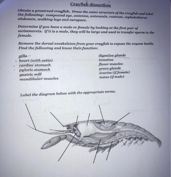
Procedure Part 1—External Anatomy of a Crayfish. 1. Put on safety goggles, gloves, and a lab apron. 2. Place a crayfish so the dorsal side is up in the dissection tray. Use the diagram below to locate the cephalothorax and the abdomen. The carapac e, a shield of chitin, covers the dorsal surface of the cephalothorax. Crayfish dissection diagrams 1. Dorsal View1. These are the crayfishs uropods. It has two pairs of these appendages.2. This is the crayfishs telson. It is used in combination with the uropods for backwards escape swimming.3. This is the crayfishs abdomen. Its paired appendages are the swimmerets and uropods.4. Start studying Crayfish dissection. Learn vocabulary, terms, and more with flashcards, games, and other study tools.
Crayfish dissection diagram. GENBIO2 - Dissection Lab - Invertebrates CRAYFISH DISSECTION Like all crustaceans, a crayfish has a fairly hard exoskeleton that covers its body. As shown in the diagram on the next page, its body is divided into two main parts, the cephalothorax and the abdomen. The cephalothorax consists of the cephalic (or head) region and the thoracic region. The part of the exoskeleton that covers the ... Crayfish Dissection Virtual Crayfish Dissection - Cornell Virtual Crayfish Dissection - Penn State By Day: Day 1 Day 2 Day 3 By Region: External Anatomy Internal Anatomy By Topic: Skeletal Integumentary Cardiovascular Muscular Endocrine Nervous Reproductive Respiratory Excretory Digestive You must create a series of labeled drawings … Continue reading "Crayfish Dissection" Use a probe to disconnect any muscle stuck to the carapace as it is lifted away. Peel the hard exoskeleton away from the underlying organs in the tail, similar.12 pages This Crayfish Dissection Guide will help students identify anatomical structures and organs of the crayfish.
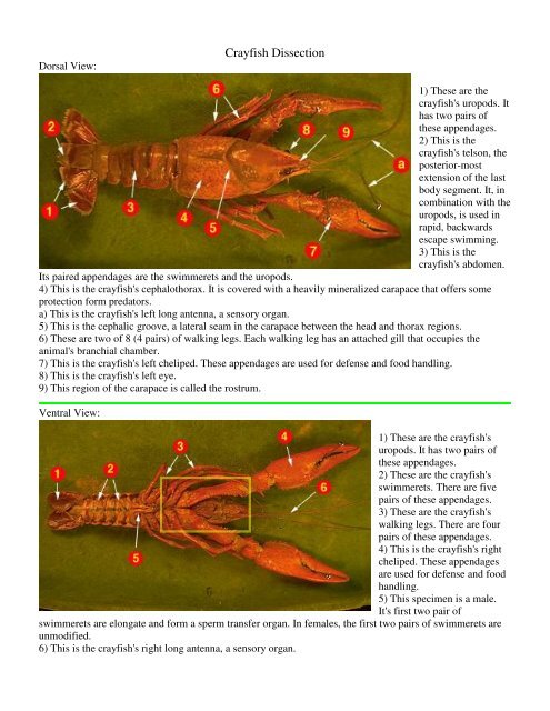
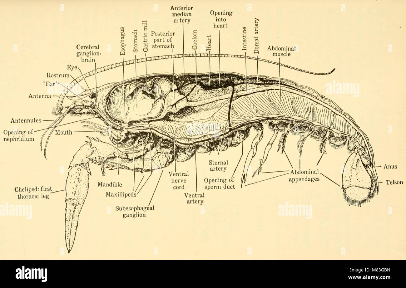
7. Between the digestive glands, you will find the small pair of white reproductive organs and cuts in the male animal. If your specimen is female, you will ... Cephalothorax. The cephalothorax as seen in the above picture makes up the crayfish's midsection. There is a tough armor that covers the vital organs and part of the head, this part of the exoskeleton is called the CARAPACE.Note how the carapace extends over the head and between the eyes, this structure is called the ROSTRUM.. Abdomen Label Crayfish External Anatomy. Read the definitions below, then label the crayfish diagram. Abdomen - The abdomen is the segmented tail area. The swimmerets, telson, and uropods are attached to the abdomen. Carapace - The protective shell (exoskeleton) of the cephalothorax. Cephalic groove - An indentation in the carapace between the head ... Shop other top-selling dissection specimens, such as earthworms for dissection, dogfish shark dissection, dissect crayfish, cow eyes, sheep brain specimen, sheep brain dissection kit, a preserved starfish, or owl pellets for classroom. ←
You may use your photocopied diagram to help you locate the organs ** Cross out the number when you have completed that step of the procedure to help you keep your place. I. Purpose The purpose of this lab is to observe the structure and function of internal organs of crayfish through dissection. II. Materials 1 crayfish 1 dissection tray Procedure Part 1—External Anatomy of a Crayfish 1. Place the crayfish with its dorsal (top) side up in a dissection tray. Use the diagram below to locate the cephalothorax and the abdomen.The carapace, a shield of chitin, covers the dorsal surface of the cephalothorax. On the carapace observe the cervical groove, an indentation, that extends across the midregion and separates the head and ... The crayfish has 8 jointed walking legs, a segmented body, 2 pairs of sensory antennae, and compound eyes. It has 2 large pincers or claws called chelipeds. If a crayfish loses a leg, the leg will regenerate (regrow). The head and thorax are fused, forming the cephalothorax. You can see the anatomy diagram of a crayfish in the following ... crayfish dissection SlideShare uses cookies to improve functionality and performance, and to provide you with relevant advertising. If you continue browsing the site, you agree to the use of cookies on this website.
Mooreschools Com
The dissection can be performed in about 30 minutes and requires only scissors, forceps, and dissecting pins. A hand lens may be helpful. Below is a brief survey of the internal and external anatomy of the earthworm. For more detailed dissection instructions and information, check out Carolina® Dissection Kits.

Crayfish Dissection Biology Junction
Jun 01, 2013 · csdn已为您找到关于carpenters postman相关内容,包含carpenters postman相关文档代码介绍、相关教程视频课程,以及相关carpenters postman问答内容。
Crayfish Dissection Ppt Video Online Download
Crayfish anatomy diagrams. Ventral view, labelled. Dorsal view. Dorsal, unlabelled. Ventral, unlabelled.Read the definitions below, then label the crayfish diagram. Abdomen - The abdomen is the segmented tail area. The swimmerets, telson, and uropods are attached to the abdomen.
Crayfish Dissection
one pair of antennules (organs of balance, touch, and taste) · one pair of longer antennae (organs of touch, taste, and smell) · one pair of mandibles (crush the ...Phylum: ArthropodaSpecies: Procambarus clarkiiGenus: ProcambarusKingdom: Animalia

Crayfish Dissection Diagram Quizlet
In this Biology lab, we will take a close look at the external and internal anatomy of a freshwater aquatic invertebrate—the crayfish. As we study the anatom...
30 Label Crayfish External Anatomy Labels For You
1. Crayfish remains go in trash can - wipe out the tray with a paper towel into the trash can first before washing tray. 2. Wash tray and tools 3. Return tray and tools to lab table Don't forget… you will be tested on the parts. Google "Crayfish dissection" for online help
Behavioral Neuroscience Crayfish Circuitry
The crayfish moves backward by forcing water forward with its tail fan. Procedure. Part 1—External Anatomy of a Crayfish. 1. Put on safety goggles, gloves, and a lab apron. 2. Place a crayfish on its dorsal side in a dissection tray. Use the diagram below to locate the cephalothorax and the abdomen.

Ppt Crayfish Dissection Laboratory Powerpoint Presentation Free Download Id 2250435
Crayfish Dissection Skills Practice Lab ... The animal shown in the diagram is a male crayfish. If your specimen is a male, locate the testis. The testis is the long, white organ under the heart and a bit forward. The sperm ducts that carry sperm from the testis open at the fifth walking leg.

Crayfish Dissection Biology Junction
Anatomy of a Shrimp/Crawfish Grade Level: 5 -12 Subject Area: Biology, Anatomy Time: Preparation: 10 minutes Activity: 3 -45 minutes Clean-up: 10 minutes Student Performance Standards (Sunshine State Standards):

Reading Arthropods Biology Ii Laboratory Manual
walking legs, Diagram Key: 1. antennae, 2. antennule, 3. cheliped, 4. eye, 5. rostrum, 6. walking legs. Like all crustaceans, a crayfish has a fairly hard exoskeleton that covers its body. As shown in the diagram on the next page, its body is divided into two main parts, the cephalothorax and the abdomen. . Label the drawing of the crayfish.

4 Schematic Illustration Of Crayfish Anatomy Showing Main Organs Download Scientific Diagram
Crayfish external anatomy diagram. Extending from the carapace is a pointy structure called the […] The sixth pair of swimmerets modified into two pairs of broad flat plates that are on the sides of the telson. External Anatomy of a Crayfish. Crayfish Dissection Crayfish Dissection Crawfish Abdomen is the main muscle that allows crayfish to swim.

New Page 1
Crayfish Dissection - Brooklynn B's Biology 11 Portfolio. CRAYFISH DISSECTION. ANALYSIS: 1. How many pairs of appendages did your crayfish have? Our crayfish had eight pairs of appendages. In total, one pair of antennae, one pair of antennules, two pairs of maxilla, three pairs of maxillipeds, and one pair of mandibles. 2. Label the diagram below.

Crayfish Dissection
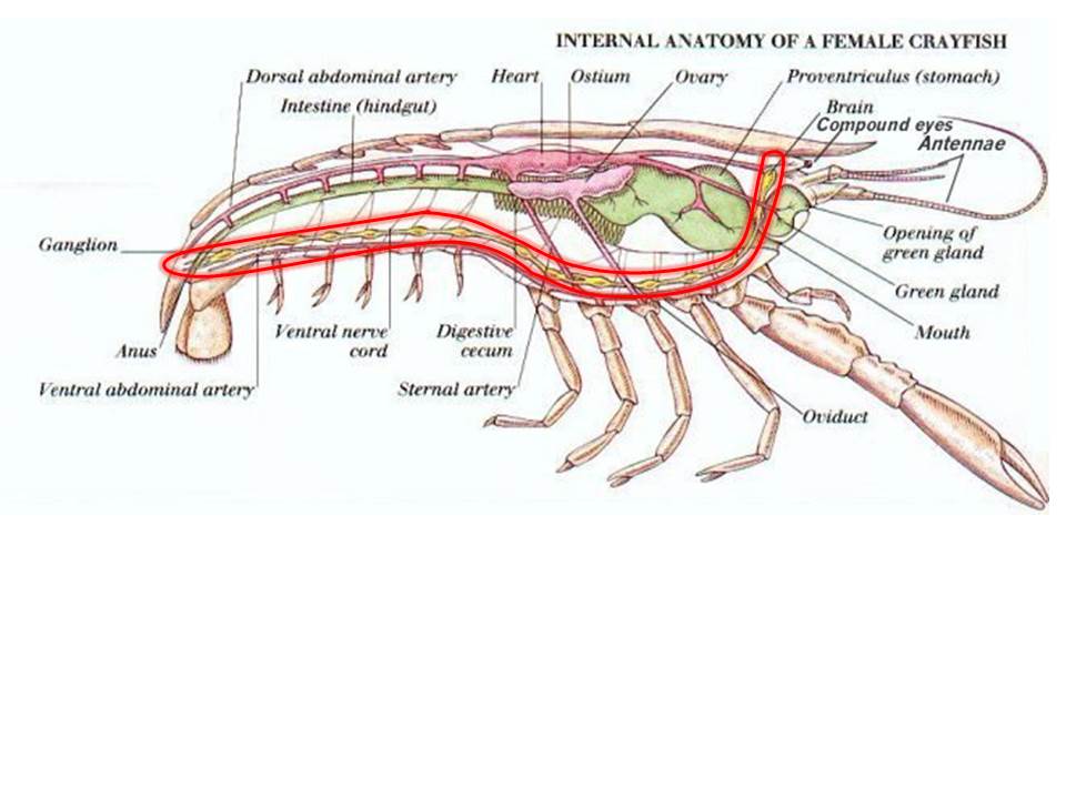
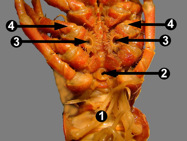
Crayfish Dissection - Internal Anatomy. INTERNAL ANATOMY: The diagram below displays the crayfish with the carapace carefully removed exposing the underlying gills and other organs. The gills, which are feather-like structures found underneath the carapace and attached to the chelipeds and walking legs, are the organs of the respiratory system ...

Crayfish Dissection Guide Crayfish Dissection Science Tools
原文地址为: 插件8:拼写检查<?php // Plug-in 8: Spell Check// This is an executable example with additional code supplied // To obtain just the plug-ins ...
Media Vwr Com
PAP Biology Dissection Test: Crayfish. abdomen. antenna. antennule. anus. section of the crayfish that contains muscles to help it swim. sensory organs used for touch, taste, and smell. sensory organs used for balance, touch, and taste. hole that expels waste.
Crayfish
Feb 23, 2004 · [prev in list] [next in list] [prev in thread] [next in thread] List: llvm-commits Subject: [llvm-commits] CVS: llvm/test/Programs/MultiSource/Benchmarks ...
Crayfish Lesson Plans Worksheets Reviewed By Teachers
substancial - Free ebook download as Text File (.txt), PDF File (.pdf) or read book online for free. contains some random words for machine learning natural language processing
Crustaceans Crayfish Dissection Naming Crayfish Kingdom Phylum Animalia
Procedure Part 1-Externa/ Anatomy of a Crayfish. 1. Put on gloves. Place a crayfish dorsal side up in a dissection tray. Use the diagram below to locate the cephalothorax and the abdomen. The carapace, a shield of chitin, covers the dorsal surface of the cephalothorax. On the carapace, observe an indentation, the

Crayfish Dissection Biology Junction
Crayfish Virtual Dissection. OBJECTIVE: Analyze images of a crayfish to study its external and internal structures and systems. Like all crustaceans, a crayfish has a fairly hard exoskel eton that covers its body. Its body is divided into two main parts, the cephalothorax and the abdomen. The cephalothorax consists of the cephalic (or head ...
Crayfish Dissection Dissection 101 Dissection Resources For Classroom Use Pbs Learningmedia
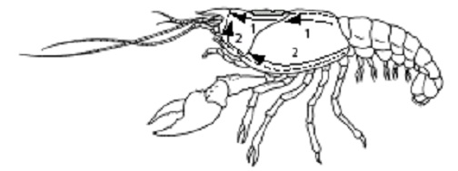
Part 2—Internal Anatomy of a Crayfish 15. Using one hand to hold the crayfish dorsal side up in the dissecting tray, use scissors to carefully cut through the back of the carapace along dissection cut line 1, as shown in the diagram below. Cut along the indentations that separate the thoracic portion of the carapace into three regions.
Plyter Com
14. $6.50. Zip. Crayfish are often one of the first specimens dissected by General Biology students. The lab worksheets provide a great introduction to animal anatomy and physiology through detailed pictures and full-color diagrams. Students will learn a great deal about the behavior and structures of a crayfish, a.
Mooreschools Com
Start studying Crayfish dissection. Learn vocabulary, terms, and more with flashcards, games, and other study tools.

Crayfish Dissection Internal Anatomy Mrs Castellucci
Crayfish dissection diagrams 1. Dorsal View1. These are the crayfishs uropods. It has two pairs of these appendages.2. This is the crayfishs telson. It is used in combination with the uropods for backwards escape swimming.3. This is the crayfishs abdomen. Its paired appendages are the swimmerets and uropods.4.

Detailed Crayfish Dissection Part I Jr High High School And College Review Youtube
Procedure Part 1—External Anatomy of a Crayfish. 1. Put on safety goggles, gloves, and a lab apron. 2. Place a crayfish so the dorsal side is up in the dissection tray. Use the diagram below to locate the cephalothorax and the abdomen. The carapac e, a shield of chitin, covers the dorsal surface of the cephalothorax.
Crayfish Dissection Mr E Science
Solved Crayfish Dissection Obtain A Preserved Crayfish Draw Chegg Com
Globetrotterscience Com
Crayfish

Crayfish Dissection Internal Anatomy Mrs Castellucci
Lab 8 Arthropods Zoo Lab Uw La Crosse
Crayfish Dissection Animal Biology 1938 18196643695 Stock Photo Alamy
Sps186 Org
1

Crayfish Dissection Diagram For Kids Pdf

Boreal Crayfish Dissection Lab Vwr

Crayfish Dissection Key Mr Lesiuk

Open Versus Closed Circulatory System Dissection Of The Crayfish And Earthworm Carolina Com
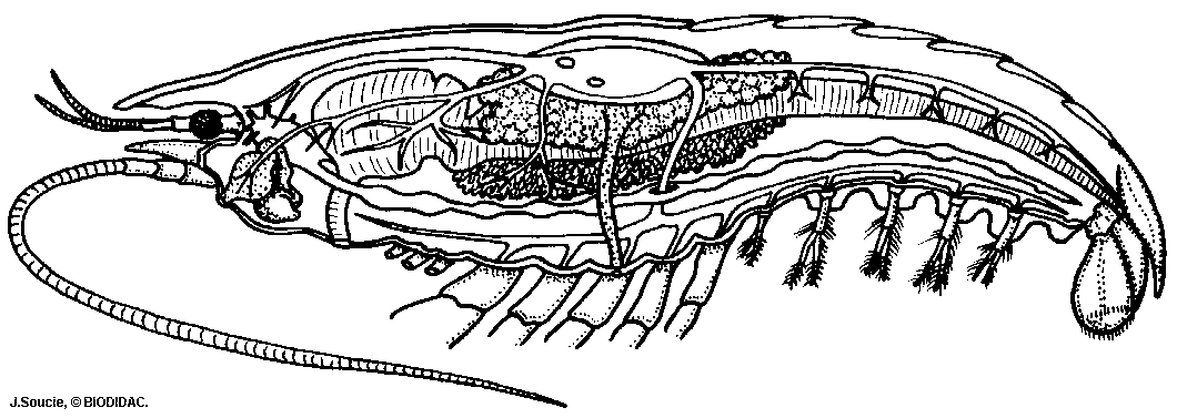
Internal Anatomy Of The Crayfish

Crayfish Anatomy Part 1 Youtube
Mooreschools Com
Comments
Post a Comment