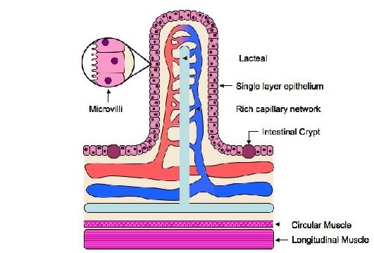
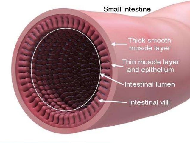
41 diagram of villus
Q. The diagram shows a tooth with signs of decay. What has made the hole in the enamel of the tooth? answer choices . acid. saliva. sugar. toothpaste. Tags: Question 6 . SURVEY . 60 seconds . Q. The diagram shows a villus. The arrows show the direction of flow within vessels associated with the villus. Which vessel carries blood to the liver ... Apr 03, 2021 · The picture above is a diagram of what is inside the villus. Source: thumbs.dreamstime.com. Ii)label the diagram of the villus below: Source: wikieducator.org. As shown in the diagram above, each villus contains a capillary bed and a after crossing the epithelium, most of these molecules diffuse into a capillary network inside the villus, and hence into.
• the wall of a villus, • the small intestine? wall of a villus small intestine A cell organ B cell organ system C tissue organ D tissue organ system 8 Phloem is an example of ... 28 The diagram shows a potato tuber that developed from the stem of a parent potato plant. Three shoots are starting to grow from the tuber. shoot

Diagram of villus
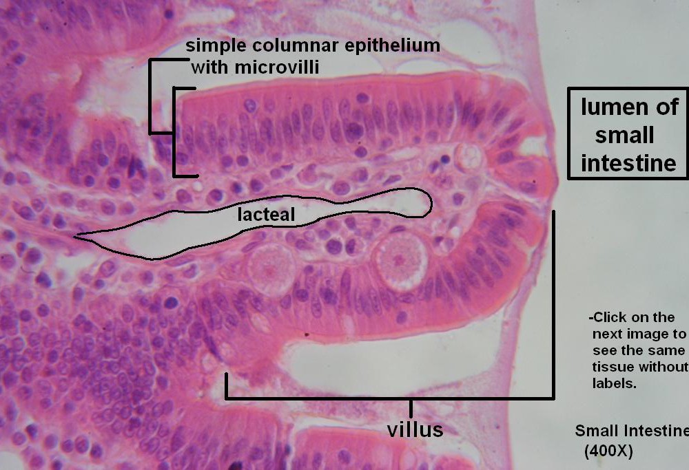
Apr 01, 2018 · This diagram represents the types of bone-marrow cell derivatives operative within the lamina propria. They include (in cerise) the subepithelial myofibroblast system (MYF); pericytes (green) supporting the subepithelial capillaries and main vasculature of the villi (artery, red: vein, blue); the lacteal (L) supported by smooth muscle cells (SM) and (purple) the muscularis mucosae (MM). Villi in the small intestine absorbs nutrients and completes the breakdown of food. Factors of its structure that help it function include. The process that the nutrients move into the villi is diffusion. The picture above is a diagram of what is inside the villus. It explains what kind of nutrients is absorbed by the blood capillary which is ... villus, plural villi, in anatomy any of the small, slender, vascular projections that increase the surface area of a membrane.Important villous membranes include the placenta and the mucous-membrane coating of the small intestine.The villi of the small intestine project into the intestinal cavity, greatly increasing the surface area for food absorption and adding digestive secretions.
Diagram of villus. Vector diagram Structure of Villi and microvilli showing arteries, veins, nerve and lymph vessel. villus stock illustrations Ciliated columnar epithelium Ciliated columnar epithelium. epithelial cells forms the lining of the stomach and intestines, duodenum, fallopian tubes, uterus, central canal of the spinal cord, nose, ears and the taste buds. villus stock illustrations Fig. 5.1 shows a longitudinal section of a villus. Fig. 5.2 shows a cross-section of the same villus at V – W. The diagrams are not drawn to the same scale. Q P W S V R cell T microvilli Fig. 5.1 Fig. 5.2 www.igexams.com Fig. 5.1 shows a longitudinal section of a villus. Fig. 5.2 shows a cross-section of the same villus at V – W. The diagrams are not drawn to the same scale. Q P W S V R cell T microvilli Fig. 5.1 Fig. 5.2 (i) Name structures P, Q, and R. 23 The diagram shows a shoot that has been placed on its side. The shoot begins to grow upwards. P Q up What causes the shoot to grow upwards? A increased cell division by meiosis at P B increased cell division by mitosis at P C more cell elongation at P than Q D more cell elongation at Q than P
15 The diagram shows a villus. Structures P and Q absorb different products of digestion. P Q Which row identifies the products absorbed by P and Q? P Q A amino acids glucose B fatty acids maltose C glucose fatty acids D maltose amino acids 16 The diagram shows a leaf attached to the stem of a plant. ... a. villus b. nephron c. ureter d. urethra. b. nephron. refer to the illustration above. the structure shown in the diagram is a(n) a. villus b. nephron c. ureter d. urethra. b. nephron. refer to the illustration above. at the location labeled "X" a. filtration is taking place Villi Diagram. Below given picture is the villi diagram: [Image will be uploaded soon] Role of Villi. The role of villi or villi function are stated below: The villi along with the microvilli support in increasing the intestinal adsorbent surface area by approximately 30-fold and 600-fold, respectively. The surface of the small intestine wall is folded, and has projections called villi. Villi is the plural of villus. The epithelial cells that cover each villus themselves have projections called ...
villus, plural villi, in anatomy any of the small, slender, vascular projections that increase the surface area of a membrane.Important villous membranes include the placenta and the mucous-membrane coating of the small intestine.The villi of the small intestine project into the intestinal cavity, greatly increasing the surface area for food absorption and adding digestive secretions. Villi in the small intestine absorbs nutrients and completes the breakdown of food. Factors of its structure that help it function include. The process that the nutrients move into the villi is diffusion. The picture above is a diagram of what is inside the villus. It explains what kind of nutrients is absorbed by the blood capillary which is ... Apr 01, 2018 · This diagram represents the types of bone-marrow cell derivatives operative within the lamina propria. They include (in cerise) the subepithelial myofibroblast system (MYF); pericytes (green) supporting the subepithelial capillaries and main vasculature of the villi (artery, red: vein, blue); the lacteal (L) supported by smooth muscle cells (SM) and (purple) the muscularis mucosae (MM).
Intestinal villus
unknown

yellow green and white map

Student Notebook Containing Notes, Diagrams and Swatches (c. 1898–1900) // Alfred Fehr (Switzerland, 1879-1955)

River City, Aerial Perspective (1979) // Bertrand Goldberg American, 1913–1997
Representation of the villus vascular system emphasizing arteriole and... | Download Scientific Diagram

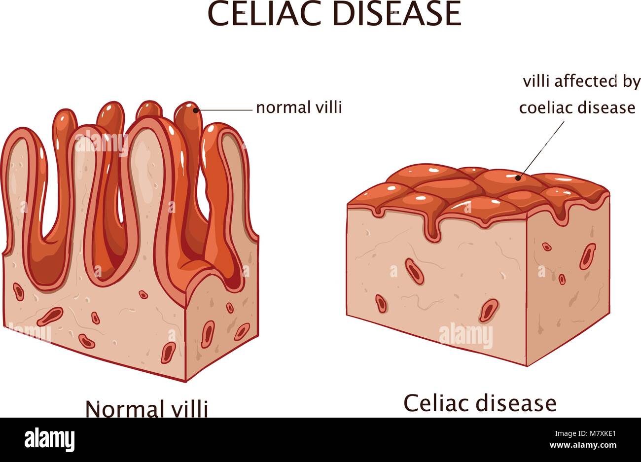
Celiac Disease. Causes, symptoms, treatment Celiac Disease
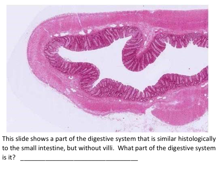
Solved: This Slide Shows A Part Of The Digestive System Th ...

Image from page 339 of "Biology of the vertebrates : a comparative study of man and his animal allies" (1949)

Differentiation markers along the crypt-villus axis in 3D ...
Gastrointestinal tract 4: anatomy and role of the jejunum and ileum | Nursing Times
Figure 1. Intestinal structure: (A) diagram of small ...
the given diagram shows the internal structure of a villus.label the parts 1 to 4.give the function of - Brainly.in

Identify the labelled parts A-D in the given figure of human
Illustration of small intestinal villi and epithelial cell turnover.... | Download Scientific Diagram

Differentiation markers along the crypt-villus axis in 3D ...
Wiring And Diagram: Diagram Small Intestine Villi

Role of villus microcirculation in intestinal absorption ...

Plan of Chicago, Chicago, Illinois, Railroad Circuits Diagram (1909) // Daniel Hudson Burnham (American, 1846-1912) Edward Herbert Bennett (American, born England, 1874-1954)
2014 WAEC Biology Theory (a) Define the following in (i) elimination (ii) excretion (iii) secretion. (b) Describe the digestion... - Myschool

unknown
AP II

Block-Neighborhood-Community: Diagram (n.d.) // Bertrand Goldberg American, 1913-1997
human heart scale model

Morphology of the intestinal villus showing neutrophil ...

Health Sciences Center, Stony Brook, New York, Sectional Diagram (c. 1974) // Bertrand Goldberg American, 1913-1997

Morphology of the intestinal villus showing neutrophil ...
Villus Diagram Diagram | Quizlet
chair parts chart hanging on wall

Villi | Definition of Villi by Webster's Online Dictionary

Immunofluorescent staining of MT1-MMP and markers for all ...

(a) Identify the parts labelled 1, 2 and 3 in the diagram ...

Image from page 391 of "The anatomy of the human body" (1844)
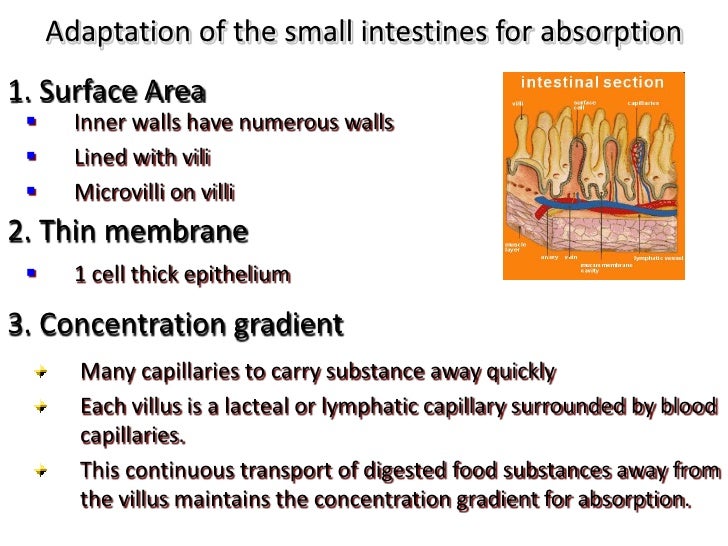
S5 Small Intestines II

Hypoxia-induced pathological changes in the small ...
Human digestive system
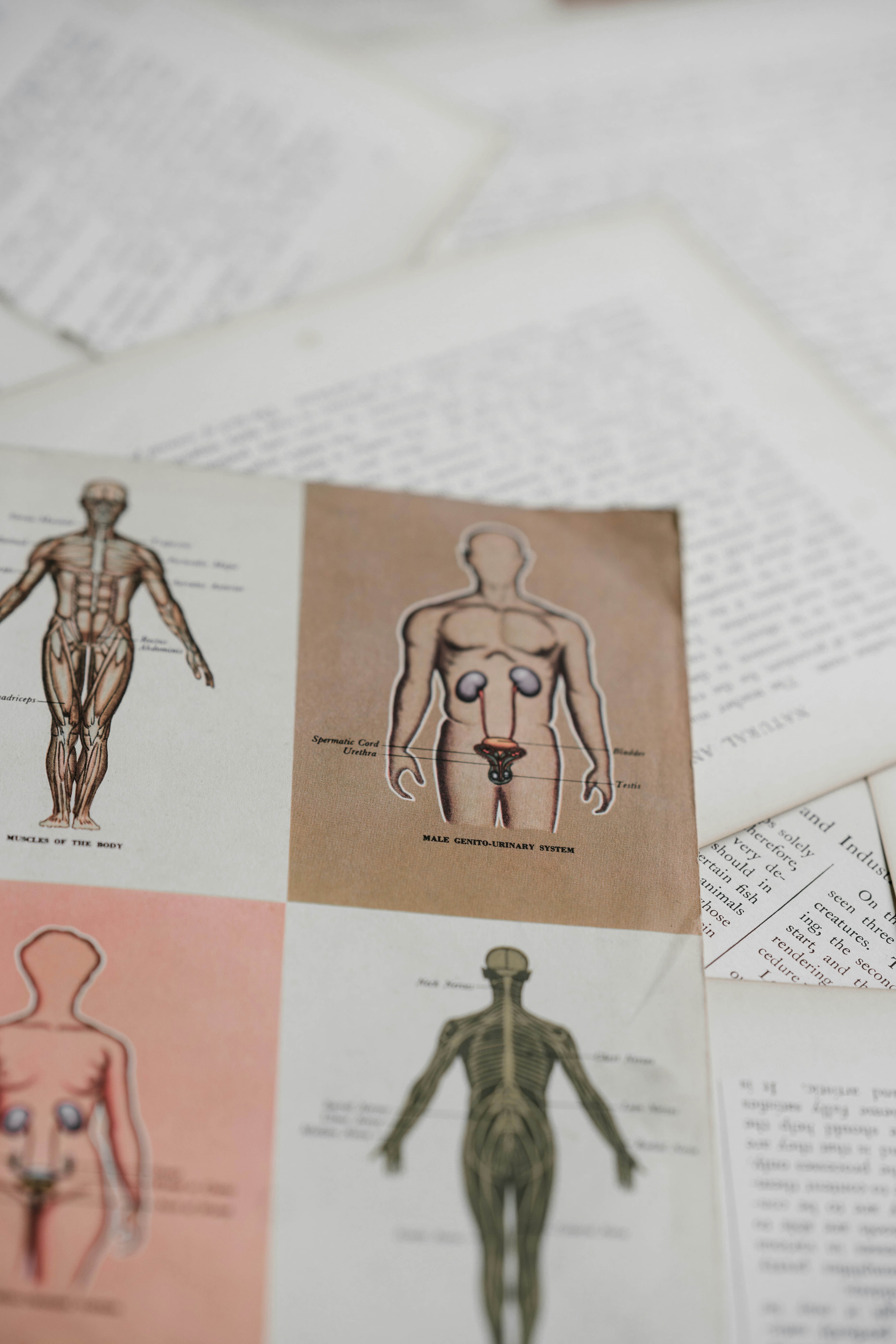
shallow focus photo of book page
Morphology of the intestinal villus showing neutrophil ...

gray rope on brown wooden table
Villi and Absorption

white printer paper with green line


Comments
Post a Comment