41 pressure ulcer sites diagram
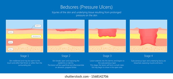
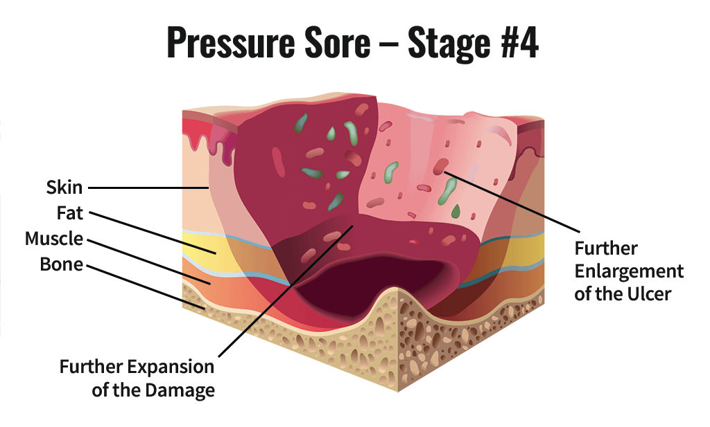
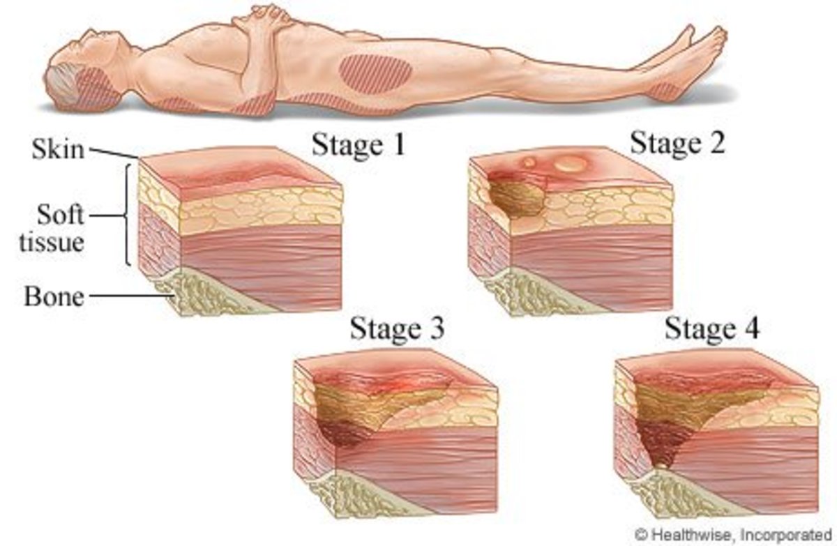
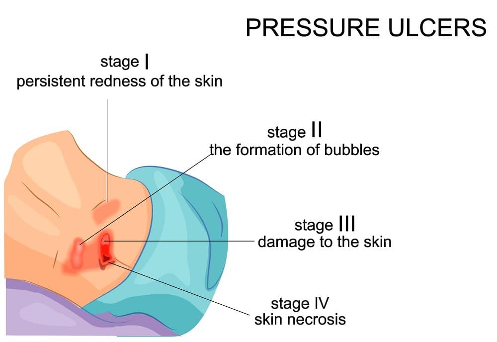
Jan 31, 2019 · Pressure ulcer risk assessment using clinical judgement alone: Braden pressure ulcer risk assessment and training: Pressure ulcer incidence Visual skin assessment Follow‐up: 8 weeks: Study population: RR 1.43 (0.77 to 2.68) 65 more per 1000 (from 35 fewer to 254 more) 180 (1 study) ⊕⊝⊝⊝ Very low 1 tissue loss this is an Unstageable Pressure Injury. + Unstageable Full-thickness skin and tissue loss in which the extent of tissue damage within the ulcer cannot be confirmed because it is obscured by slough or eschar. If slough or eschar is removed, a Stage 3 or Stage 4 pressure injury will be revealed.
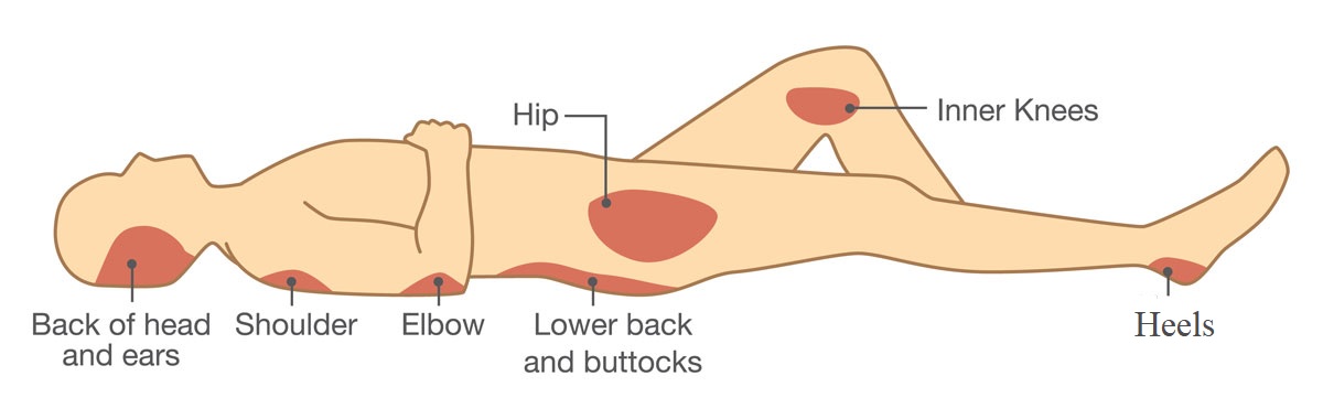
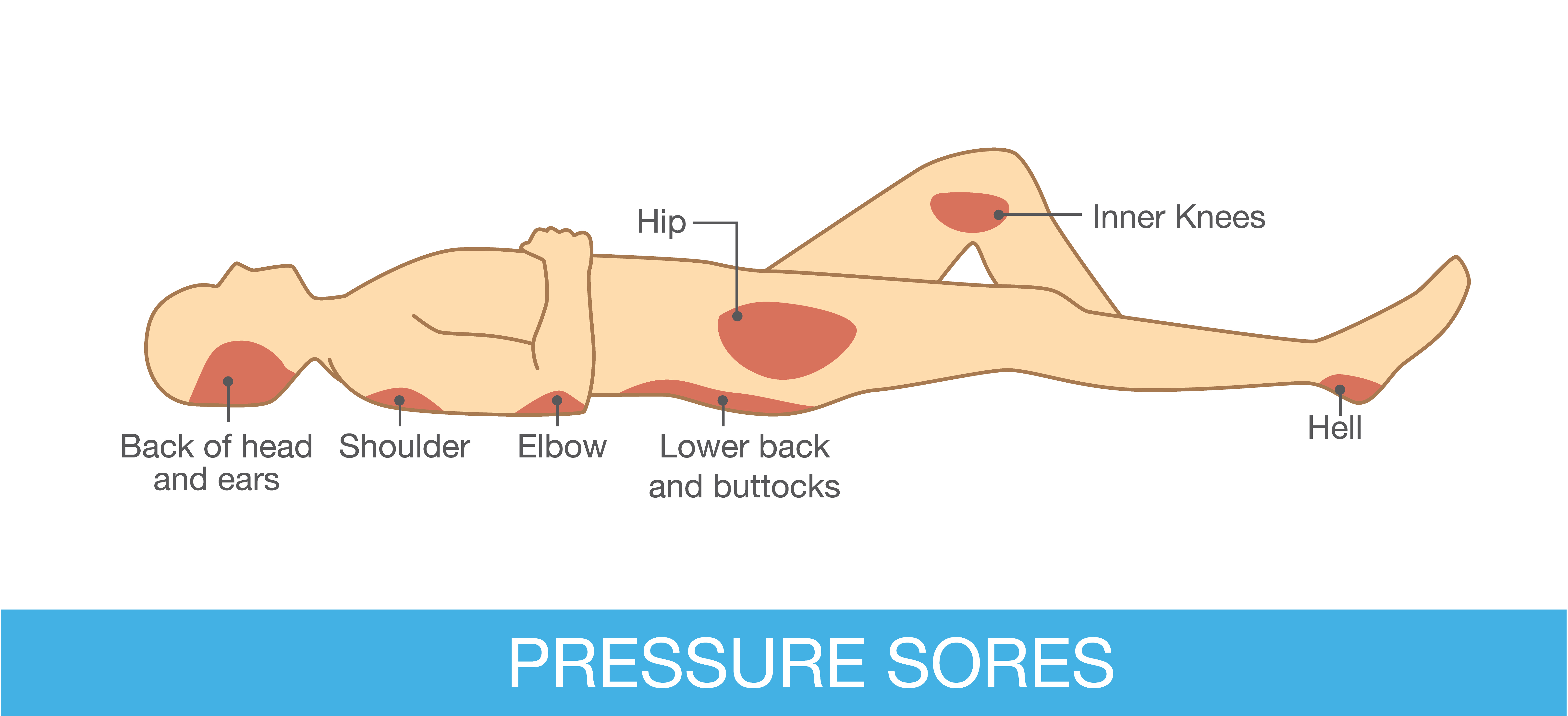
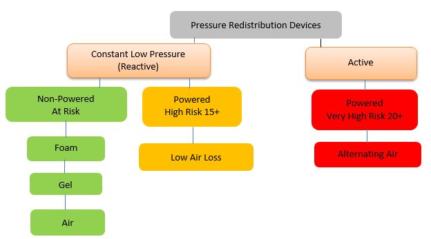
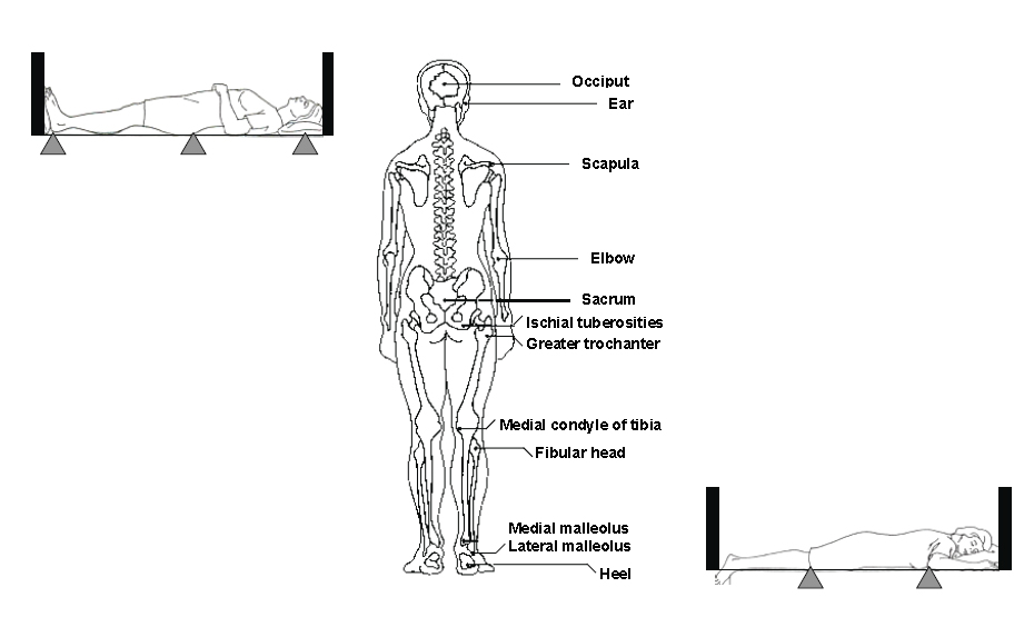
Authorship. Areas where bones are close to the surface (called "bony prominences") and areas that are under the most pressure are at greatest risk for developing pressure sores. In bed, body parts can be padded with pillows or foam to keep bony prominences (areas where bones are close to the skin surface) free of pressure.

Pressure ulcer sites diagram
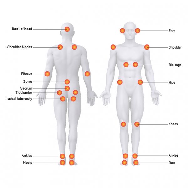
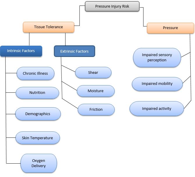
Pressure ulcers, also known as pressure sores or bed sores, are localised damage to the skin and/or underlying tissue that usually occur over a bony prominence as a result of usually long-term pressure, or pressure in combination with shear or friction. The most common sites are the skin overlying the sacrum, coccyx, heels, and hips, though other sites can be affected, such as the elbows ... Pressure Ulcers Reference: Stephanie Amlung, PhD, RN; Wendy L. Miller, and Linda M Bosley, BSM "The 1999 National Pressure Ulcer Prevalence Survey: A Benchmarking Approach," Advances in Skin & Wound Care; 14(6):297-301, Nov-Dec 2001. Pressure injuries on the mucous membranes present and are staged differently from cutaneous pressure ulcers, and they are usually attributed to a medical device or tube. Nasogastric or orogastric tubes, oxygen cannulas or masks, endotracheal tubes, and urinary and fecal containment devices pose a risk of causing local ischemia to tissue in the ...
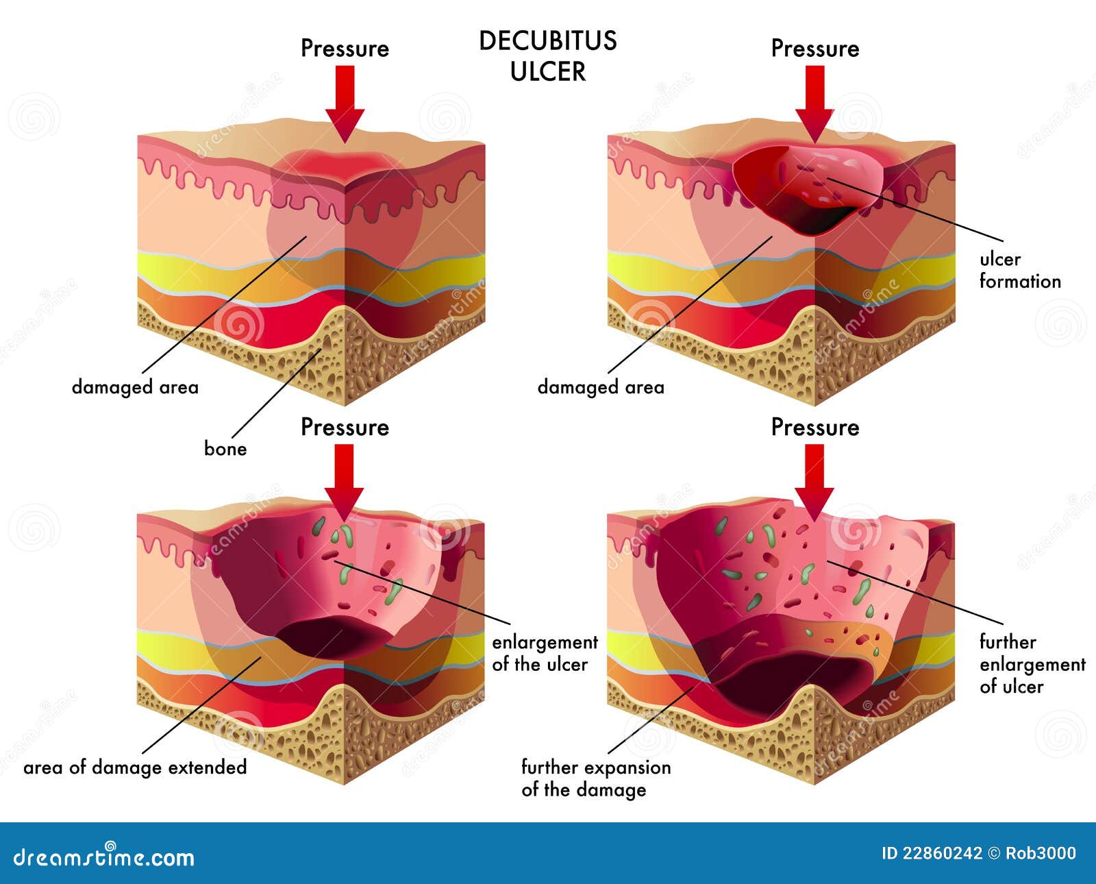
Pressure ulcer sites diagram. Pressure ulcers (bedsores) Factsheet 512. LP. September 2016. Pressure ulcers - also called pressure sores or bedsores - can develop if someone spends too long sitting or lying in one position. They are a . particular risk for people with dementia. It is important for anyone caring for a person with dementia to know about . pressure ulcers. –Pressure ulcer –Rash –Infection, cellulitis •Deficiencies can also affect skin: –Vitamin C deficiency causes purplish blotches on lightly traumatized areas. –Zinc deficiency causes redness of the nasolabial fold and eyebrows. 17 Pressure ulcers are the third most expensive disorder after cancer and cardiovascular diseases. In Japanese Geriatric Health Services facility, the immobile geriatric patients represent 91% of total population with pressure ulcer in the Geriatric Health Service facility. The incidence of pressure ulcers is different in each clinical setting. Pressure sores (also known as decubitus ulcers, bedsores, and pressure ulcers) are areas of skin and tissue that have been damaged by inadequate blood flow because of sustained pressure. Nursing home residents and hospital patients are at increased risk for pressure ulcers because of decreased mobility, wheelchairs, and bed confinement.
Each year, more than 2.5 million people in the United States develop pressure ulcers. These skin lesions bring pain, associated risk for serious infection, and increased health care utilization. The aim of this toolkit is to assist hospital staff in implementing effective pressure ulcer prevention practices through an interdisciplinary approach to care. • pressure from equipment being used to monitor or treat an individual (eg urinary catheter, oxygen tubing). The most common sites for pressure ulcers to develop are: • the sacrum (the curved triangular bone just above the buttocks) - accounts for over 30% of all pressure ulcers; • the heels - accounts for 25-30%; pressure ulcer ( Flowchart) Use Creately's easy online diagram editor to edit this diagram, collaborate with others and export results to multiple image formats. We were unable to load the diagram. You can edit this template and create your own diagram. Creately diagrams can be exported and added to Word, PPT (powerpoint), Excel, Visio or any ... Common Sites for Pressure Ulcers. Create healthcare diagrams like this example called Common Sites for Pressure Ulcers in minutes with SmartDraw. SmartDraw includes 1000s of professional healthcare and anatomy chart templates that you can modify and make your own.
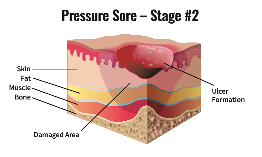
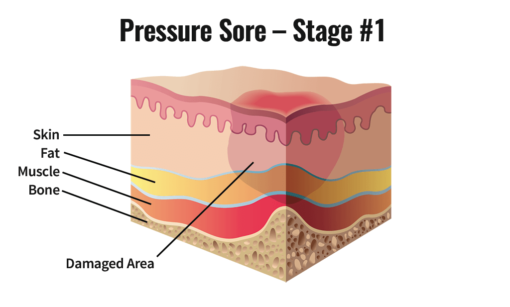
These sites tend to develop pressure ulcers more quickly as these are bony areas, and the fat that works as natural cushion is less in these areas. But pressure ulcers are said to be largely preventable with the help of protective devices and proper management. There are custom designed gel and pneumatic wheelchair cushions which are easily ... trochanters, all of which are common sites for pressure ulcer development. 2 This coding and identifi cation prob-lem became critically important when the Centers for Medicaid & Medicare Services began to deny payment for pressure ulcers (PU) that occur while a patient is hospital-ized or being treated in a long-term care facility. 3 Understand Pressure Injury Staging, Braden Scale scoring, and Braden Sub score For all inpatients: Inspect and monitor skin (at least daily) and as clinically indicated: Nursing documentation for any pre-existing wounds can be found in "Wound/Ulcer Assessment" tab of the "Wound Care Intake/Management Tool" Feb 01, 2018 · Top diagram showing pressure ulcers Stage I: skin intact. Stage II: partial skin loss. Stage III: full-thickness skin loss, subcutaneous tissue exposed. Stage IV: muscle, tendon, bone or organs exposed. Bottom diagram showing unstageable pressure ulcer with tissue damage hidden from observer by eschar over entire wound. Deep tissue injury ...
Where Do Pressure Areas Develop Pressure Ulcer Basics For Aged Care Workers
The chart contains diagrams of the body and the location of a pressure ulcer is recorded by numbering the location of each pressure ulcer and recording its severity in a table below. Use this grading tool to accurately assess the grade of each pressure ulcer. It works well with the Scottish Adapted European Pressure Ulcer Advisory Panel (EPUAP ...
Los Angeles Nursing Home Bed Sores Lawyer Pressure Ulcers
Secondary hypertension is hypertension due to an identifiable cause, and may result in certain specific additional signs and symptoms. For example, as well as causing high blood pressure, Cushing's syndrome frequently causes truncal obesity, glucose intolerance, moon face, a hump of fat behind the neck and shoulders (referred to as a buffalo hump), and purple abdominal stretch marks.
Pressure Ulcer Images Stock Photos Vectors Shutterstock
A pressure ulcer is a localized injury to the skin and/or underlying tissue, usually over a bony prominence, as a result of pressure, or pressure in combination with shear. (NPUAP-EPUAP, 2009) ... Source for diagram: Saha S, Smith MEB, Totten A, et al Gov. document ...

Immunomodulation As Rescue For Chronic Atonic Skin Wounds Trends In Immunology
What are the common sites for pressure ulceration? The areas that are particularly prone to pressure sores are those that cover the bony areas such as: Heels. Ankles. Pelvis, especially over the tailbone (sacrum) Hipbones (ischium) Shoulders or shoulder blades (scapula) Backs or sides of the knees. Toes.

Sarawak Hospice Society Pressure Sore Bed Sore
Common Locations for Pressure Sores. Pressure Injury Prevention prevention. For people who use a wheelchair, pressure sores often occur on skin over the following sites: Tailbone or buttocks. Shoulder blades and spine. Backs of arms and legs where they rest against the chair. For people who are confined to a bed, (home or nursing home) common ...

What Is A Sacral Pressure Ulcer Nursing Home Abuse Center
A pressure ulcer is an ulcer related to some form of pressure and should not be confused with ulcers relating to disease (like cancer), vascular flow (venous or arterial) or neuropathy (like in persons with diabetes) You should be able to see a “cause and effect” relating to pressure with the ulcer.

Jaypeedigital Ebook Reader
Despite 2014 and subsequent updates to the ICD-10, there are still no specific anatomical coding designations for the coccyx, ischial tuberosities, the posterior iliac crests, or the trochanters, all of which are common sites for pressure ulcer development.2 This coding and identification problem became critically important when the Centers for ...

Stages Of Pressure Ulcers Diagram Quizlet
vanGilder C, Amlung S, Harrison P, Meyer S. Results of the 2008-2009 International Pressure Ulcer Prevalence™ Survey and a 3-year, acute care, unit-specific analysis. Ostomy Wound Management, 2009;55(11):39-45. 19 . Objectives- Participants will: • Differentiate. pressure ulcers from other skin injuries •Describe pressure ulcer . stages ...
Plos One Neuroprotective Effect Of Erythropoietin Against Pressure Ulcer In A Mouse Model Of Small Fiber Neuropathy
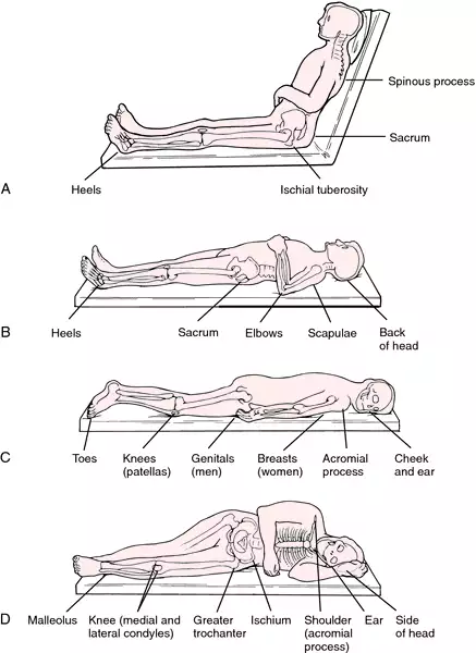
guidelines, Assessment and Prevention of Pressure Ulcers and Assessment and Management of Stage I - IV Pressure Ulcers. The purpose of this learning package is to assist health care providers in long-term care facilities to gain the knowledge and skill required to manage the unique challenges inherent in the positioning of residents with varying

File Pressure Ulcer Sites Png Wikimedia Commons
1 Assessment Chart for Wound Management For multiple wounds complete formal wound assessment for each wound. Add Inserts as needed. Factors which could delay healing: (Please tick relevant box) Immobility Poor Nutrition Diabetes Incontinence Respiratory / Circulatory
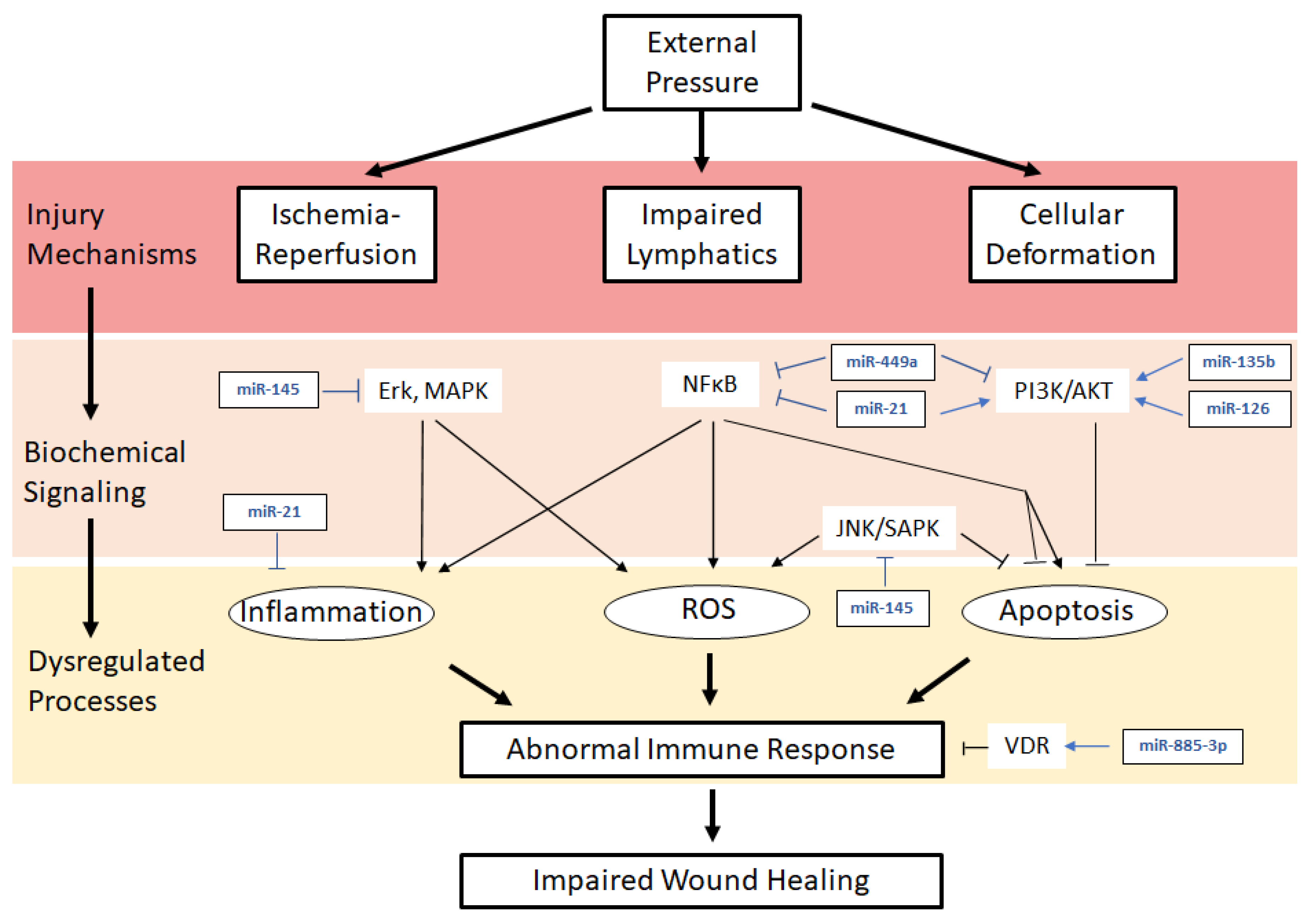
Ijms Free Full Text Role Of Micrornas In Pressure Ulcer Immune Response Pathogenesis And Treatment Html
notification from [HCP name] when the client is discharged from service for treatment to pressure ulcer(s) so that consultation with Nursing Services regarding any ongoing care needs to pressure ulcer(s) or skin may occur. A non-professional is providing skin care (treatment) for a client who has a pressure ulcer.
Pressure Wounds And Ulcers Inspirational Stories
Cost and prevalence of pressure ulcers Development of the SKIN Bundle and how it can be used to help prevent pressure ulcers How the SKIN Bundle was implemented at Cardiff and Vale University Health Board Keywords: Pressure ulcer/SKIN Bundle/ Prevention This article has been double-blind peer reviewed Nursing Practice Innovation Pressure ulcers

Prevention Of Pressure Ulcers Among Individuals Cared For In The Prone Position Lessons For The Covid 19 Emergency Journal Of Wound Care
2009 Pressure Ulcer Definition "… localized injury to the skin and/or underlying tissue usually over a bony prominence, as a result of pressure, or pressure in combination with shear." 12 NPUAP/EPUAP Pressure Ulcer Prevention and Treatment Guidelines.
Skin Pressure Sores After Spinal Cord Injury
of developing pressure injuries and residents with exiting PIs. Research shows that a resident at risk for skin breakdown can develop a PI within hours of onset of pressure, and interventions should be implemented promptly to prevent PIs. Federal regulation lists examples of resident risk factors for pressure injuries/ulcers 1:

Common Locations For Pressure Ulcer Formation Over 95 Of Pressure Download Scientific Diagram
Acquired Pressure Ulcers (HAPU) since 2013. The run chart shows sustained improvement from September 2016. New solutions were required to address remaining wards with high levels of HAPU. One option was a handheld device to detect early signs of skin damage. There was evidence to support its use,

Pressure Ulcer Sites Fn 2 Unit 10 Diagram Quizlet
The depth of a Category/Stage III pressure ulcer varies by anatomical location. The bridge of the nose, ear, occiput and malleolus do not have (adipose) subcutaneous tissue and Category/Stage III ulcers can be shallow. In contrast, areas of significant adiposity can develop extremely deep Category/Stage III pressure ulcers.

Pdf Pressure Ulcers Impact Etiology And Classifi Cation Semantic Scholar
Pressure injuries on the mucous membranes present and are staged differently from cutaneous pressure ulcers, and they are usually attributed to a medical device or tube. Nasogastric or orogastric tubes, oxygen cannulas or masks, endotracheal tubes, and urinary and fecal containment devices pose a risk of causing local ischemia to tissue in the ...
1
Pressure Ulcers Reference: Stephanie Amlung, PhD, RN; Wendy L. Miller, and Linda M Bosley, BSM "The 1999 National Pressure Ulcer Prevalence Survey: A Benchmarking Approach," Advances in Skin & Wound Care; 14(6):297-301, Nov-Dec 2001.
Development Of Pressure Ulcers Causes And Signs Scottish Acquired Brain Injury Network E Learning
Pressure ulcers, also known as pressure sores or bed sores, are localised damage to the skin and/or underlying tissue that usually occur over a bony prominence as a result of usually long-term pressure, or pressure in combination with shear or friction. The most common sites are the skin overlying the sacrum, coccyx, heels, and hips, though other sites can be affected, such as the elbows ...
Skin Pressure Sores After Spinal Cord Injury
Pressure Ulcers Cause Owlcation
Clinical Guidelines Nursing Pressure Injury Prevention And Management

Elderly Abuse In Nursing Homes Jerome Salmi Kopis Llc
World Wide Pressure Ulcer Prevention Day November 21 2013
Pressure Ulcers Prevalence Brigham And Women S Hospital

St Christopher S Pressure Ulcers St Christopher S
Cyberounds Cme

Allevyn Life For Pressure Ulcer Prevention Smith Nephew Corporate

Pressure Ulcer Lhsc
Pressure Ulcer Stock Illustrations 74 Pressure Ulcer Stock Illustrations Vectors Clipart Dreamstime

Pressure Sores Prone Areas Affected By Body Position On Bed A Download Scientific Diagram
Skin Pressure Sores After Spinal Cord Injury

Proper Positioning For The Prevention Of Pressure Sores And Muscle Contracture

Pressure Sore Locations Diagram Stock Image C030 6113 Science Photo Library

Pdf Pressure Ulcers Impact Etiology And Classifi Cation Semantic Scholar
Pressure Ulcer Definition Of Pressure Ulcer By Medical Dictionary

Skeleton Presentation Common Sites Of Pressure Ulcers The Most Common Download Scientific Diagram
Clinical Guidelines Nursing Pressure Injury Prevention And Management

Pressure Ulcer Text Images Music Video Glogster Edu Interactive Multimedia Posters
1
What Is A Pressure Sore Distasio Law Firm


Comments
Post a Comment