40 frog label diagram
Microsoft Word - Labeled Diagram for Lab #19 - Frog Dissection.doc Author: CoteJ Created Date: 7/19/2012 3:17:13 PM ... The nasal openings, are also call EXTERNAL NARES, found toward the tip of the snout will closes when the frog is under water. Label the mouth, tympanum, and the external nares on Figure 1. 5. Feel the frog's skin. It is smooth, moist and thin. The frog can breathe directly through its skin as well as with its lungs.
#frog #frogdiagram #howtodrawStudents need to learn about the basic parts of a frog. So in this video, I try to help you with drawing a labeled diagram of a ...

Frog label diagram
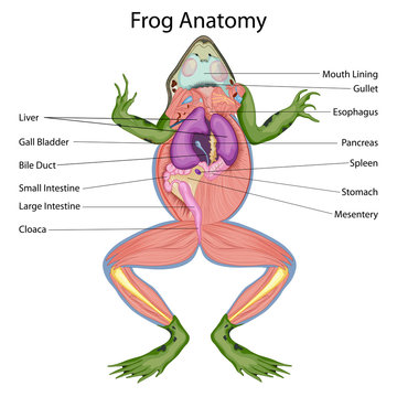
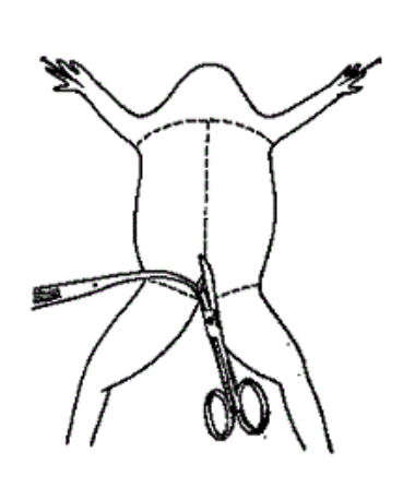
Internal Frog Anatomy. Lay the frog ventral surface up on the dissecting tray. Pinch the loose skin at the center of the frog's stomach and make an initial cut with the scissors into the skin. Cut through the skin, following the pattern shown in the diagram below. Follow the same pattern to cut through the muscle and reveal the internal organs. 25 Easy Frog and Toad Ideas and Activities Teaching Frog and Toad will feel simple with these 25 ideas and activities. You'll find science and reading sources about frogs and toads, the life cycle of a frog, frog facts and a frog craft or two to round it all off. Flamingo Diagram Label a diagram of a flamingo. Answers: Forest Animals in English A Label Me! Printout Label the fox, squirrel, deer, antler, bear, claw, raccoon, hedgehog, mouse, and worm in English. Answers: Frog Life Cycle Diagram Label a diagram of the frog's life cycle. Answers
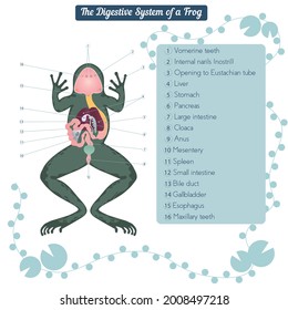
Frog label diagram. << Back to Frog Activities Click on the image below to see it in its own window (close that window to return to this screen) OR Right click and save image to your hard drive to print from your own image software at your convenience. The major organs involved in the process of digestion in frogs include mouth, pharynx, esophagus, stomach, small intestine, large intestine, and cloaca. Accessory organs such as the liver, pancreas, and gallbladder are also an important part of the digestive system of frogs. This BiologyWise post provides a labeled frog digestive system diagram to help you understand the digestive process in ... Frog Body Parts and Functions External Anatomy of the Frog External Anatomy of the Frog Determine if your frog is a Male or Female The sex of a frog may be determined externally by examining the thumb pads on the front feet. The thumb pads of males are enlarged at the base as in the drawing on the right. please help me out! i need to do well on this to pass the course. grade 10. please create diagram of frog to label; Question: please help me out! i need to do well on this to pass the course. grade 10. please create diagram of frog to label
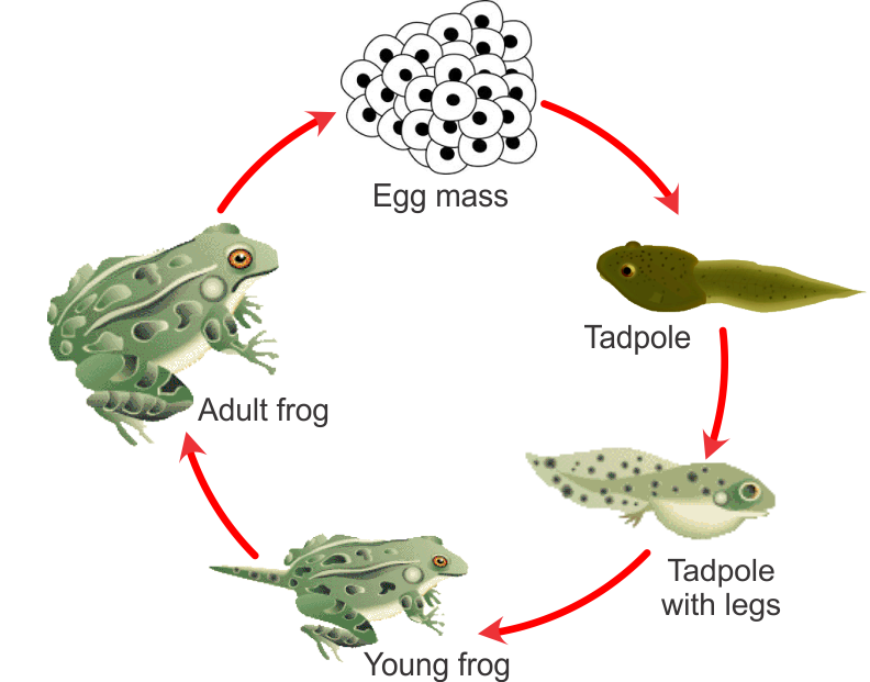
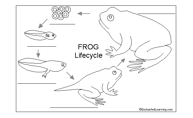
Ventral view of muscles in a frog. A diagram of all the key muscle groups within the Frog and how they help the Frog function day to day. Ventral view of muscles in a frog. A diagram of all the key muscle groups within the Frog and how they help the Frog function day to day. This frog dissection page describes how to dissect a frog and lists organs for students to find and check as the perform the dissection. It covers digestive system and urogenital system and contains several pictures for students to label. Mar 03, 2012 · In the mean time we talk about Frog Anatomy Labeling Worksheet, we have collected particular related photos to complete your references. frog internal anatomy diagram, frog dissection worksheet answer key and labeled frog anatomy diagram are three of main things we want to present to you based on the gallery title. Read the definitions below, then label the frog life cycle diagram. egg - Tiny frog eggs are laid in masses in the water by a female frog. The eggs hatch into tadpoles. tadpole - (also called the polliwog) This stage hatches from the egg. The tadpole spends its time swimming in the water, eating and growing.
Frog Dissection Pre-lab Directions: Watch the virtual Dissection ... “Introduction” & “External Anatomy” to answer the questions. Use the second web link to label the frog’s internal organs with location and function. Expect to take about an hour to prepare for this dissection. 1. Phylum_____ 2. SubPhylum _____ ... of the different systems of the frog. Label the diagram in Part 2 of the questions section. Refer to the diagram of the internal structures of the frog if necessary. Digestive system: 12. When the frog ingests its food, it passes along the esophagus to the stomach. From the stomach, the food passes through the small intestines, through a short ... Frog Dissection Pre-lab. Directions: Watch the virtual Dissection “Introduction” & “External Anatomy” to answer the questions. Use the second web link to label the frog’s internal organs with location and function. Expect to take about an hour to prepare for this dissection. Start studying frog urogenital system. Learn vocabulary, terms, and more with flashcards, games, and other study tools.
Draw a labelled diagram of the life history of frog and ...
1 of 2. One labeled diagram and one blank diagram for students to complete. diagram amphibian animal frog body anchor chart chart.
draw a labeled diagram of the various stages in the lifecycle ...
Frog Dissection Diagram and Labeling. High Resolution PDF for Printing. Click Here. Citing Research References. When you research information you must cite the reference. Citing for websites is different from citing from books, magazines and periodicals. The style of citing shown here is from the MLA Style Citations (Modern Language Association).

draw labelled diagram of life history of frog and identify ...
the Frog •Nictitating Membrane - A transparent part of a frog’s lower eyelid that moves over the eye to clean it and protect it. •Cloacal Opening - Opening of cloaca through which undigested food, urine, eggs, and sperm are passed. •Vocal Sacs - The vocal sac is the flexible membrane of skin possessed by most male frogs.
Frog Life Cycle Diagram N3 free image download
A drawing of the frog showing the major organs, drag the names of the organs to the label. This quiz is self grading and intended for practice.
Frog dissection Images, Stock Photos & Vectors | Shutterstock
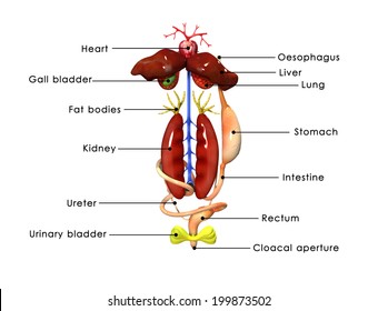
In frog the sexes are separate. The urinogenital organs can be studied under the following heads: 1. Excretory System: The excretory system in both male and female frog is similar. The excretion is mainly carried out with the help of a pair of kidneys, a pair of ureters, a urinary bladder and cloaca.

draw labelled diagram of life history of frog and identify ...
Label the Anatomy of the Frog ©Sheri Amsel www.exploringnature.org Anatomy of the Frog lungs liver gall bladder fat bodies kidney small intestine mesentery common iliac artery femoral artery sciatic artery conus arteriosus of heart stomach pancreas spleen bladder esophagus carotid artery aortic arch ...
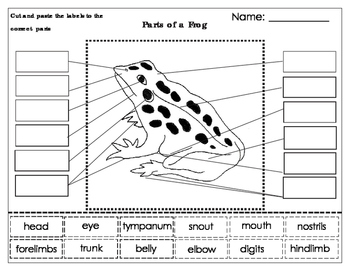
Label parts of an Amphibian:Frog
The digestive system of the frog includes the alimentary canal (alimentary tract) and the various associated digestive glands.. Digestive System of Frog. The alimentary canal of the frog is complete. It is a long, coiled tube of varying diameter that extends from mouth to cloacal aperture of the frog.

33 Diagram Of Frog With Label - Labels Design Ideas 2020
In this you are going to learn how to draw labelled diagram of Frog easily for the classification of animals of Kingdom Animalia and phylum chordata.#frogdia...
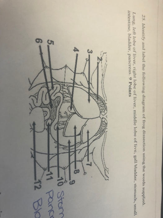
Solved 25. Identify and label the following diagram of frog ...
Diagram 1: External Features 1 1. BEFORE DISSECTION. Draw the frog's front foot and the back foot on the diagram. Remember, there is a difference in a frog's front and back feet. You will write in the length of these legs during the lab. 2. BEFORE DISSECTION. Label the following on Diagram 1: front leg, hind
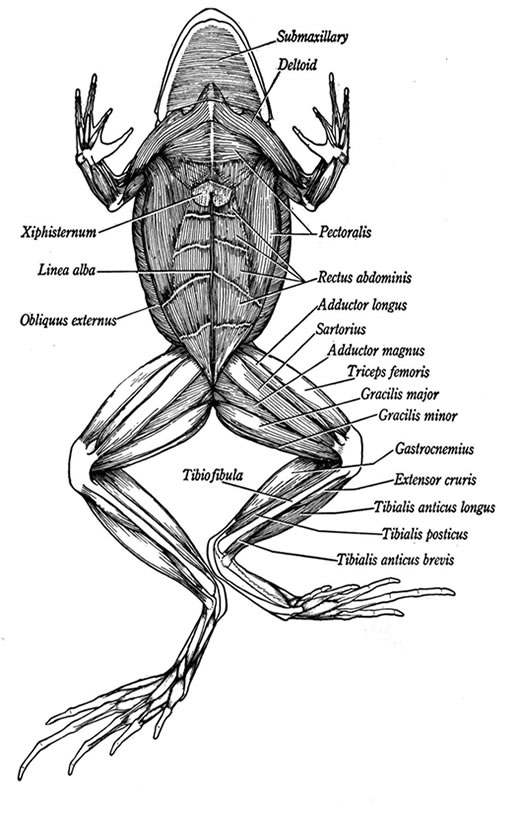
Ventral View Of Muscles In A Frog - Anatomy Diagram
Male frogs have bean-shaped testes attached to their kidneys. When you're done, print out this diagram and fill in the labels yourself to test your knowledge of frog anatomy: Diagram of dissected frog to label (.pdf)
313 BEST Frog Dissection IMAGES, STOCK PHOTOS & VECTORS ...
Flamingo Diagram Label a diagram of a flamingo. Answers: Forest Animals in English A Label Me! Printout Label the fox, squirrel, deer, antler, bear, claw, raccoon, hedgehog, mouse, and worm in English. Answers: Frog Life Cycle Diagram Label a diagram of the frog's life cycle. Answers
Lab Frog Dissection Internal Part II
25 Easy Frog and Toad Ideas and Activities Teaching Frog and Toad will feel simple with these 25 ideas and activities. You'll find science and reading sources about frogs and toads, the life cycle of a frog, frog facts and a frog craft or two to round it all off.

Diagram Of External Features Of Frog || Labelled Diagram Of ...
Internal Frog Anatomy. Lay the frog ventral surface up on the dissecting tray. Pinch the loose skin at the center of the frog's stomach and make an initial cut with the scissors into the skin. Cut through the skin, following the pattern shown in the diagram below. Follow the same pattern to cut through the muscle and reveal the internal organs.
Week 2: Frog and Butterfly Life Cycles - "At Last"
Frog anatomy Images, Stock Photos & Vectors | Shutterstock

SMARTboard Frog Activities - Frog Diagram - Label the Parts ...
Frogs in the Classroom

Frog anatomy labeled vector illustration scheme. educational ...

Parts of a Frog Diagram (freebie) | Frog, Frogs for kids ...

Poison Arrow Frogs Printout- Enchanted Learning Software

Frog (Pickerel) Labeling Page

Frog Skeleton Labeling (pt 1) Diagram | Quizlet

LLA BIOLOGY: Simple Frog Diagram

How To Draw A Frog Very Simple & Easy | Labelled Diagram Of Frog | Biology Diagram

Diagram Of Internal Organs Of Frog || Labelled Diagram Of Frog || Class 11 || Biology
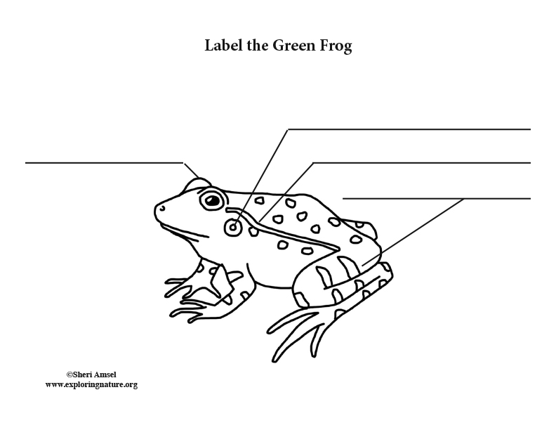
Frog (Green) Labeling Page

Search "Frogs" & "Printouts" - Enchanted Learning
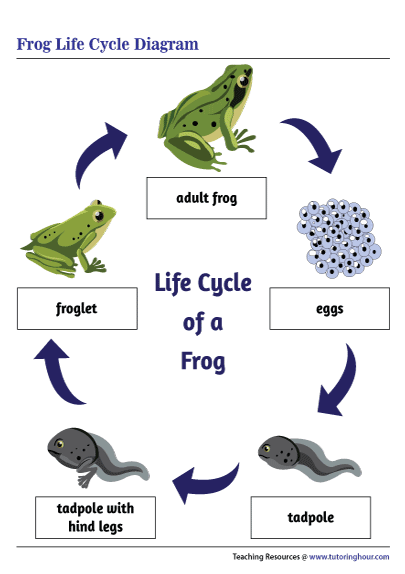
Printable Frog Life Cycle Diagram
Frog life cycle - Teaching resources

Draw a neat and well labelled diagram of male reproductive ...

Frog Dissection: External and Internal - Biology LibreTexts
Frog Dissection: External and Internal - Biology LibreTexts
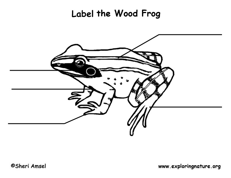
Wood Frog Labeling Page
Diagrams: Frog Dissection
.PNG)
Draw a suitable diagram of external feature of frog
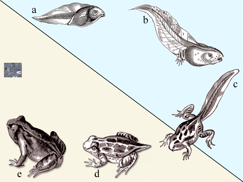
Life Cycle of the Frog

How to Draw Life Cycle of a Frog Diagram !

Labelled Diagram Of Frog

Frog Life Cycle Labelling Worksheet
Frog development examples (article) | Khan Academy


Comments
Post a Comment