41 arm veins diagram
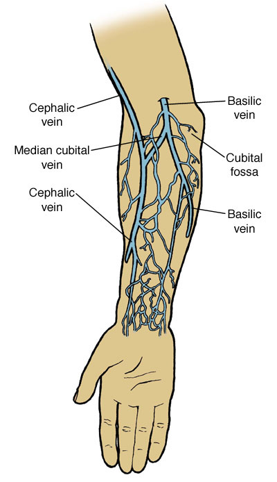
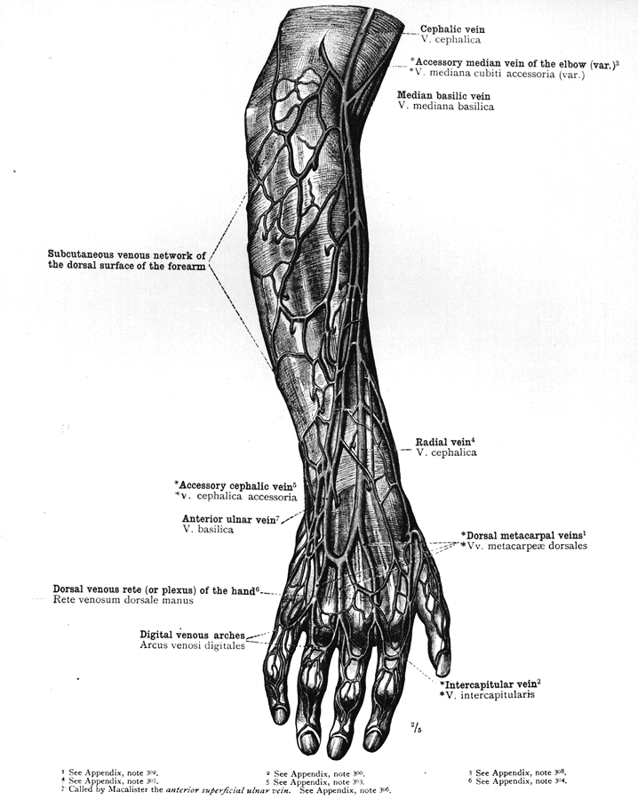
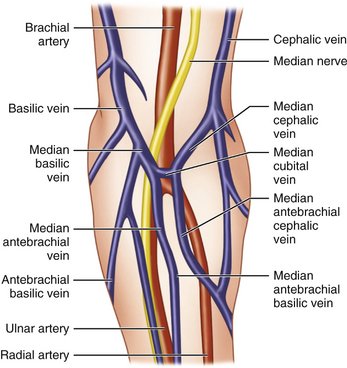
Feb 11, 2021 · The basilic vein is a superficial vein that drains the medial aspect of the arm and empties into the brachial vein proximally. Median cubital vein. The basilic and cephalic veins are joined at the anterior aspect of the elbow by the median cubital vein, often chosen as the site for blood removal for the purpose of blood testing. Accessory Cephalic Vein: this vein comes off the cephalic vein (hence its name) and is easy to stabilize. It is relatively large so this vein can hold an 18 gauge and easily a 20 or 22 gauge IV. Tip: when going for this vein for an IV go below the bend of the arm rather than in the bend. This vein extends down below the bend.
Jul 18, 2016 · The external jugular vein (v. jugularis externa) (Figs. 12-1 though 12-6, 12-8, and 12-10) is the main channel for return of venous blood from the head.It begins by the union of the linguofacial and maxillary veins, caudal to the mandibular salivary gland or at a transverse plane through the cricoid cartilage and the axis.
Arm veins diagram
May 11, 2018 · A bulging hand vein could be the result of a blood clot deep in the veins of the arm. Hand diagram. Use this interactive 3-D diagram to explore the hand. Treatment for prominent hand veins. The arm veins would be last exam-ined with this approach. Knowledge of the normal spectral waveform in the upper extremity vein is essential in the exam-ination of these veins (Figure 4). The spectral Doppler signals are characterized by 2 phasic variations in amplitude. Cardiac pulsatility man- Jan 05, 2019 · Grab the patient's lower arm (below site of puncture) firmly to draw the. Phlebotomists are considered to have occupational exposure to blood borne pathogens. . Although the larger and fuller median cubital and cephalic veins of the arm . The diagram below indicates in green the proper area to use for heel. Arm Veins For Venipuncture .
Arm veins diagram. Major Arm Veins. Here are a number of highest rated Major Arm Veins pictures on internet. We identified it from honorable source. Its submitted by presidency in the best field. We allow this nice of Major Arm Veins graphic could possibly be the most trending subject with we allocation it in google benefit or facebook. Venipuncture Sites Diagram. Adhesive bandages / tape - protects the venipuncture site after collection. .. The diagram below indicates in green the proper area to use for heel punctures for. Oct 13, Phlebotomy, the practice of drawing blood from a vein, is a proficiency that After identifying the site for the blood draw, gather the appropriate. Cannulation Diagram (Right Arm) ... using concomitant ulnar artery and ulnar vein or concomitant radial artery and radial vein in patients with minimum artery and vein diameters of 2.0 mm at the fistula creation site who have chronic kidney disease and need hemodialysis. ... Ensure the patient's arm is restrained to minimize movement during ... Diagram. Explore the interactive 3-D diagram below to learn more about the arm. Anatomy and function of arm nerves. ... Each arm contains several important veins and arteries. Veins carry blood ...
Although the larger and fuller median cubital and cephalic veins of the arm . The diagram below indicates in green the proper area to use for heel. Knowledge of vein selection, the order of draw, test-specific handling, default to the cephalic vein on the lateral or thumb side of the arm as a. GREAT diagram on upper extremity venipuncture sites! Start studying Antecubital veins of the arm. Learn vocabulary, terms, and more with flashcards, games, and other study tools. Dec 21, 2021 · Arteries, veins and nerves of the arm (a diagram) The main artery of the upper limb is the axillary artery–it is a continuation of the subclavian artery. The axillary artery continues down the arm as the brachial artery, then splits into the ulnar and radial arteries in the forearm. Browse 2,961 veins and arteries diagram stock illustrations and vector graphics available royalty-free, or start a new search to explore more great stock images and vector art. The circulatory or cardiovascular human body system medical illustration.
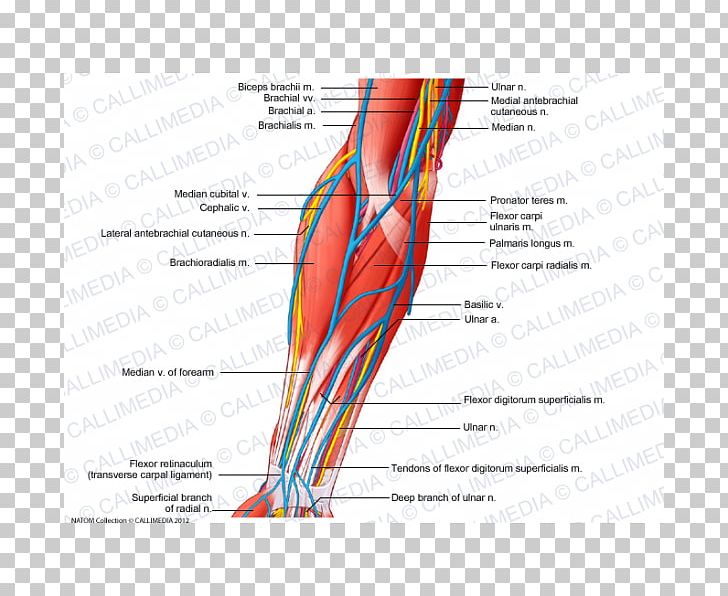
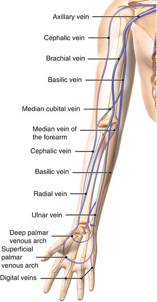
Visit Arm Veins For Venipuncture. This chapter covers all the steps recommended for safe phlebotomy and The diagram in Section , shows common positions of the vessels, but many DO NOT insert the needle where veins are diverting, because this increases the.The global leader in vein visualization schematron.orge Today · Reduce Costs · Vein ... The subclavian vein is a continuation of the axillary vein, which is located under the arm. The subclavian vein extends along the medial (middle) side of a muscle called the anterior scalene muscle. From there, the subclavian vein continues to the outer border of the first rib where it then joins the internal jugular veinto form the brachiocephalic vein (also called the innominate vein). Start studying arteries and veins of arm. Learn vocabulary, terms, and more with flashcards, games, and other study tools. The vessels of the arms are part of the circulatory system, which provides nutrients to the tissues. The arteries deliver freshly oxygenated blood to muscles and bone. The veins return oxygen ...
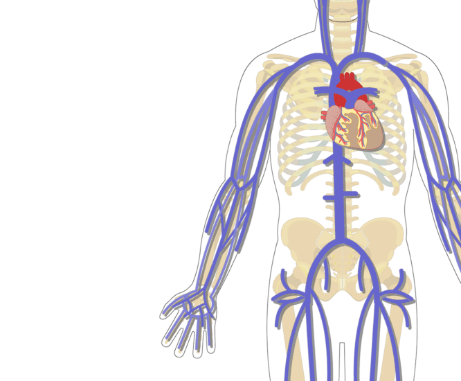
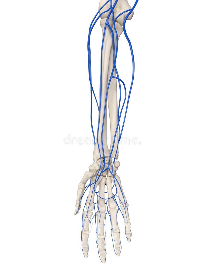
The Major Systemic Veins
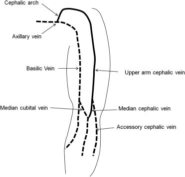
Venipuncture Module 1_Anatomy Of The Arm And Vein Location Venipuncture Module 1_Anatomy of the Arm and Vein Location - Download as PDF File .pdf), Text file .txt) or read online. The placement of these three veins forms a letter "H" as in the diagram below. there is no distinct branch from the cephalic vein to the basilic vein.
![Arm: Anatomy [+video] - Lecturio Medical](https://cdn.lecturio.com/assets/Arteries-of-the-arm.png)
Arm: Anatomy [+video] - Lecturio Medical
Diagram of all veins in arm. Posted Feb 15, 2005. by luvrn. Register to Comment. I was wondering if anyone knows of a site on the web that lists all the veins of the arm. Not just the bascilic and cephalic veins. Any vein that we put an I.V. in we have to document what vein it was inserted in. I can find the major ones but not all the ...

review labtest2 - veins arm Diagram | Quizlet
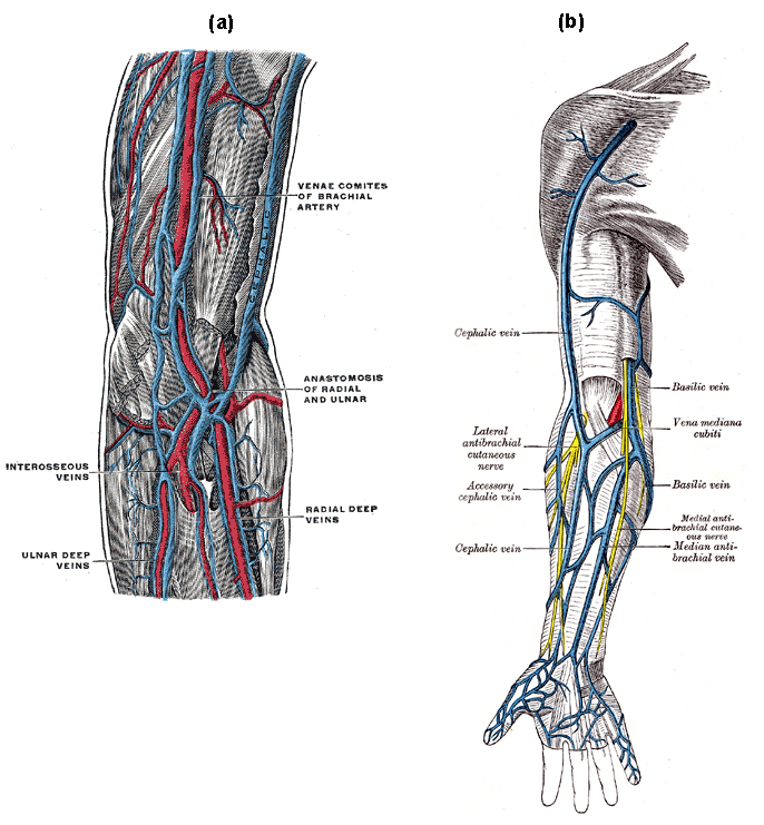
From the ulnar side (medial side of forearm) the basilic vein runs up the postaxial border of the upper limb, pierces the deep fascia halfway between the elbow and axilla, and joins the brachial veins to form the axillary veins. A third important superficial vein of the forearm is the median forearm vein (or median antebrachial vein ).

Vector illustration of a veins of the arm Stock Vector Image ...
Arteries and Veins of the Arm. Create healthcare diagrams like this example called Arteries and Veins of the Arm in minutes with SmartDraw. SmartDraw includes 1000s of professional healthcare and anatomy chart templates that you can modify and make your own. 13/71 EXAMPLES. EDIT THIS EXAMPLE. CLICK TO EDIT THIS EXAMPLE.
The arm anatomy stock illustration. Illustration of vein ...
Anal veins (A1, A2, A3) - unbranched veins behind the cubitus The costa (C) is the leading marginal vein on most insects, although a small vein, the precosta, is sometimes found above the costa. In almost all extant insects, [1] : 41–42 the precosta is fused with the costa; the costa rarely ever branches because it is at the leading edge ...

arm vein diagram - Google Search | Arm veins, Veins ...
Diagram of veins in arm for phlebotomy. A dictionary file. dict_files/eng_com.dic This class can parse, analyze words and interprets sentences. It takes an English sentence and breaks it into words to determine if it is a phrase or a clause. It can also counts the total number of words in a sentence, checks if a word is a palindrome and can ...

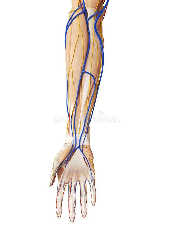
Anatomy of the Nerves, Arteries and Veins of the Arm (Upper ...
Sep 20, 2017 - The Axillary Vein This large vessel lies on the medial side of the axillary artery. It fully overlaps the artery anteriorly while the arm is abducted. The axillary vein, the extension of the basilic vein, begins at the inferior border of the teres major muscle. It ends laterally to the first rib, where it turns […]
Anatomy Atlases: Anatomy of First Aid: A Case Study Approach ...
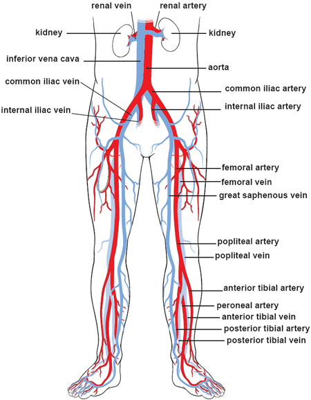
Jul 29, 2020 · There are three major types of blood vessels: arteries, capillaries and veins. Blood vessels are often named after either the region of the body through which they carry blood or for nearby structures. For example, the brachiocephalic artery carries blood into the brachial (arm) and cephalic (head) regions. One of its branches, the subclavian ...
Venous Drainage of the Upper Limb - Basilic - Cephalic ...
3. How to Draw the Artery and Veins Diagram As the cardiovascular system is complex, drawing arteries and veins may seem difficult. If the students follow the step-by-step method, they can make a cardiovascular system connected with arteries, veins, and capillaries. The student can opt for freehand drawing to create a diagram of arteries and veins.

Unlabelled Image Of The Major Systemic Veins Of The - Arm ...
Jan 05, 2019 · Grab the patient's lower arm (below site of puncture) firmly to draw the. Phlebotomists are considered to have occupational exposure to blood borne pathogens. . Although the larger and fuller median cubital and cephalic veins of the arm . The diagram below indicates in green the proper area to use for heel. Arm Veins For Venipuncture .

1,102 Arm Veins Photos and Premium High Res Pictures - Getty ...
The arm veins would be last exam-ined with this approach. Knowledge of the normal spectral waveform in the upper extremity vein is essential in the exam-ination of these veins (Figure 4). The spectral Doppler signals are characterized by 2 phasic variations in amplitude. Cardiac pulsatility man-

Veins of arm Diagram | Quizlet
May 11, 2018 · A bulging hand vein could be the result of a blood clot deep in the veins of the arm. Hand diagram. Use this interactive 3-D diagram to explore the hand. Treatment for prominent hand veins.

Median Vein Of Forearm Diagram

Vein Pelvis Abdomen Human anatomy, arm, hand, people png | PNGEgg

Arm veins Diagram | Quizlet

Forearm Vein Access for Radial Procedures: An Easy Method for ...
Arm Veins Stock Illustrations – 128 Arm Veins Stock ...
Anatomy Atlases: Illustrated Encyclopedia of Human Anatomic ...
Anterior Compartment Of The Forearm Nerve Muscle Vein PNG ...
Circulatory Routes | Boundless Anatomy and Physiology
23: Anatomy for Venipuncture | Pocket Dentistry

major veins of arm Diagram | Quizlet

Upper limb anatomy | Superficial veins, Upper limb anatomy ...
![Arm: Anatomy [+video] - Lecturio Medical](https://cdn.lecturio.com/assets/Veins-of-the-arm.png)
Arm: Anatomy [+video] - Lecturio Medical
Vascular Anatomy Of The Upper Limb

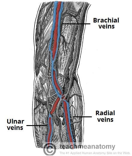
Deep veins of the upper extremity - UpToDate
Ultrasound Anatomy of Peripheral Veins and Ultrasound-Guided ...

Superficial Veins of Upper Limb - Basilic & Cephalic veins | Anatomy Tutorial

Median Cubital & Antebrachial Veins: Locations & Functions ...

Blood vessels of the arm and hand - Anatomy and Physiology

Human Anatomy Scientific Illustrations Arm Veins High-Res ...

1 Veins of the upper arm (used with permission N. Moureau ...
Illustrations of the Blood Vessels

Thumb Elbow Vein Forearm Anatomy, hand, angle, hand, anatomy ...

Arteries and veins of the upper limb | Sketchy Medicine
Superficial Veins of Forearm : Mnemonic | Epomedicine
Upper limb: Arteries, veins and nerves | Kenhub

Arm Veins Anatomy - Anatomical Charts & Posters
Anatomy and Physiology of the Arm

Basilic vein | Radiology Reference Article | Radiopaedia.org

C Blood Vessels - Dental Anatomy - Global Healthcare


Comments
Post a Comment