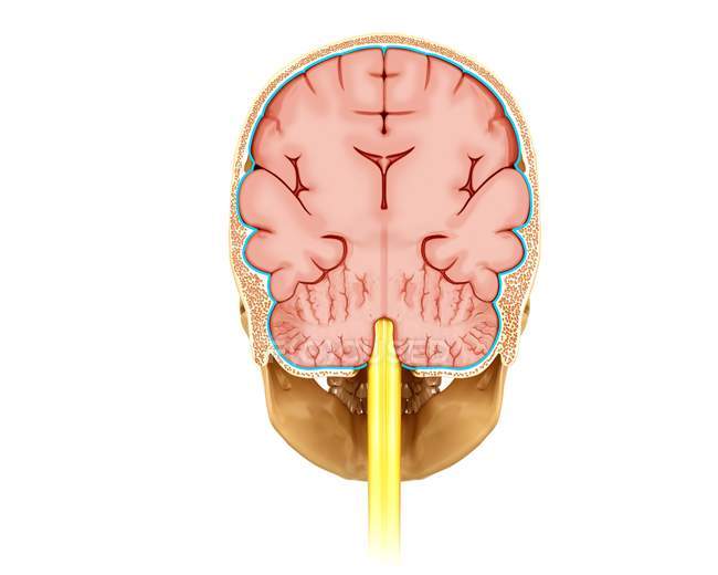
0 brain cross section diagram
Mid Sagittal Cross Section Of Brain Brain Anatomy Brain Anatomy And Function Brain Science. Median Section Of Human Brain Anatomical Structure Diagram Infographic Chart With All Parts Cerebellum Thalamus Human Brain Diagram Brain Anatomy Brain Diagram. Genu of corpus callosum (cross section) Lateral ventricle. The superior portions of the lateral ventricle can be seen in the midline. The frontal horn (occupying the frontal lobe) and body (extending into the parietal lobe) of the right and left lateral ventricles can be seen separated anteroposteriorly by the thin band of septum pellucidum.The ventricles are home to the choroid plexus, which ...
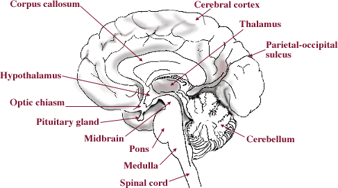
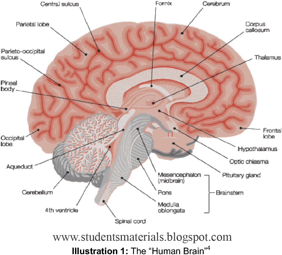
Figure 4: Mid-sagittal section of brain showing diencephalon (includes corpus callosum, fornix, and anterior commissure) Marieb & Hoehn (Human Anatomy and Physiology, 9th ed.) - Figure 12.10 Exercise 2: Utilize the model of the human brain to locate the following structures / landmarks for the diencephalon: Thalamus Intermediate mass

Brain cross section diagram
Anatomy of the Brain Chart 20x26. The 20" x 26" (51 x 66 cm) chart beautifully illustrates cerebral hemispheres, lobes of the brain, cross section of meninges and venous sinuses, typical nerve cell, synapses, typical glial cells, arteries of the brain, vagus nerves, circulation of cerebrospinal fluid, midbrain with corpus callosum, sagittal ... Brain cross section diagram. Calculate the length of cb Joe tapped his maple tree to get syrup. A vintage wall art print featuring illustrated art on an antique background. Graphic Of Brain Showing The Flow Of Information To The Thalamus Cerebral Cortex Amygdala Hippocampus Spinal Locus Coeruleus Cerebral Cortex Constellations. Cross Section Brain Diagram Brain Diagram Human Brain Nervous System Problems Human Brain Diagram Labeled Unlabled And Blank Brain Diagram Human Brain Diagram Human Brain Vertical Section Of A Human Brain Showing The Medulla Pons Cerebellum Hypothalamus Thalamus Midbrain Stock Vector Human Brain Human Brain Diagram Brain Brain Anatomy Easy To Edit Illustration Of Brain […]
Brain cross section diagram. spinal cord cross section, 5x shows: gray matter (golden butterfly), white matter, central canal, dorsal & ventral root, dorsal root ganglion, meninges, dorsal horn, ventral (anterior) horn, & anterior horn cells (motor neuron cell bodies) - cross section of the brain stock pictures, royalty-free photos & images. Regions of the Brain; Interactive Modules; Videos; Cross-Sections; MRIs; 3D Resources; Stroke Model; Neuroanatomy Syllabus Find Cross section of brain stock images in HD and millions of other royalty-free stock photos, illustrations and vectors in the Shutterstock collection. Thousands of new, high-quality pictures added every day. Find Brain Cross Section Diagram stock video, 4k footage, and other HD footage from iStock. Great video footage that you won't find anywhere else.
Dementia is caused when the brain is damaged by diseases, such as Alzheimer's disease or a series of strokes. Alzheimer's disease is the most common cause of dementia, but not the only one. A person with dementia will experience symptoms depending on the parts of the brain that are damaged, and the disease that is causing the dementia. Hacking, C. Brainstem cross-sectional anatomy (diagrams). Case study, Radiopaedia.org. (accessed on 18 Jan 2022) . Loading images... Axial sections of the brainstem with major nuclei and tracts. Brain Cross section. STUDY. Learn. Flashcards. Write. Spell. Test. PLAY. Match. Gravity. Created by. Scrux PLUS. Key Concepts: Terms in this set (11) Cerebrum. Area of the brain responsible for all voluntary activities of the body. Corpus Colllosum. the large band of neural fibers connecting the two brain hemispheres and carrying messages ... MRI Atlas of the Brain. This page presents a comprehensive series of labeled axial, sagittal and coronal images from a normal human brain magnetic resonance imaging exam. This MRI brain cross-sectional anatomy tool serves as a reference atlas to guide radiologists and researchers in the accurate identification of the brain structures.
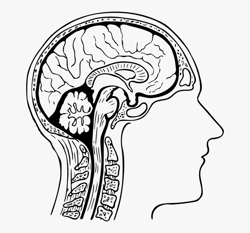
1] Brain Transection Levels-- relates tranverse sections to major divisions of the whole brain; particularly helpful for students tasked with assembling a series of brain transection images into correct rostro-caudal order. 2] Brain Transection Atlas-- useful to identify components within images of brain transections. Two modes are featured: 1) focus on names and see the components labeled and described, or 2) focus on components and see the names high-lighted. Cross Section Brain Diagram Brain Diagram Human Brain Nervous System Problems. Cross Section Showing Deeper Structure Brain Anatomy Medical Anatomy Human Anatomy And Physiology. Parts of the corpus callosum the genu and the posterior body can be appreciated in the midline of the section. We will start with a cross section of the head, where the different structures of the brain are visible. The brain is part of the central nervous system responsible for various functions, ranging from simple homeostasis to higher cognitive functions like critical thinking, memory etc. Cross section diagram of brain. The brain which is housed and protected by in the bones of the skull makes up all parts of the central nervous system above the spinal cord. Cross Section Of Human Eye Eps8 Human Brain Anatomy Human Brain Diagram Brain Diagram Brain Parts To Know Brain Diagram Brain Anatomy Human Brain Anatomy I Pinimg Com ...
cross section brain Diagram | Quizlet
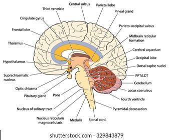
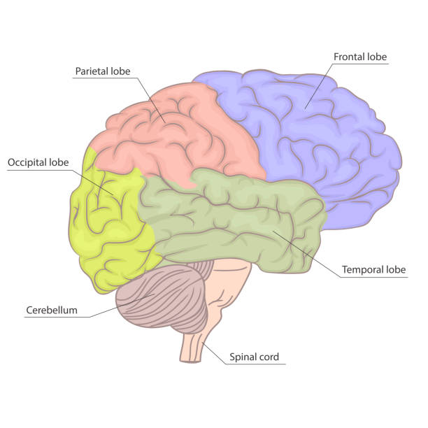
Important sulci of the cerebrum include the central or Rolandic sulcus, the lateral or Sylvian sulcus, the parieto-occipital sulcus and the Calcarine sulcus. These sulci help to divide the cerebrum into its four lobes. Given below is a diagram of brain, its functions, detailing the four lobes and their associated structures. Four Lobes of the Brain

Anatomy of Human Brain. Medical Diagram of the Structure of ...
Choose from Brain Cross Section Diagram stock illustrations from iStock. Find high-quality royalty-free vector images that you won't find anywhere else.
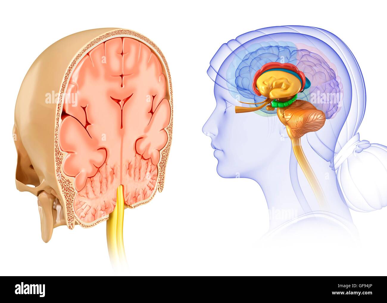
Illustration of human brain cross-section Stock Photo - Alamy
Spinal Cord - Cross-Sectional Anatomy. Start Quiz Want to learn faster? Look no further than these interactive, exam-style anatomy quizzes. Learn anatomy ... Parts of the Brain Quiz. Test your knowledge with the parts of the brain and their functions in a fun and interactive way. Click and start your quiz immediately!

Human Brain Anatomy Infographic Diagram Stock Vector ...
This is an online quiz called Cross-section Brain Labeling There is a printable worksheet available for download here so you can take the quiz with pen and paper. Your Skills & Rank
Horizontal sections of the brain: Anatomy | Kenhub
Cross sectional anatomy: MRI of the brain. An MRI was performed on a healthy subject, with several acquisitions with different weightings: spin-echo T1, T2 and FLAIR, T2 gradient-echo, diffusion, and T1 after gadolinium injection. We obtained 24 axial slices of the normal brain.

Human brain anatomy diagram cross section with all lobes and ...
The procedure is divided into three main sections: Examination of the Exterior of the Brain, Examination of the Mid-Sagittal Plane of the Brain, Examination of two Frontal Cuts. Examination of the Exterior of the Brain. The first portion of the dissection will be a detailed examination of the brain surface.
Human brain cross-section — brain structure, cross section ...
Find the perfect drawing diagram cross section stock photo. Huge collection, amazing choice, 100+ million high quality, affordable RF and RM images. No need to register, buy now!

Mid-Sagittal Cross Section of Brain | Brain anatomy, Brain ...
Jun 11, 2018 - Brain Cross Section Diagram Blank - See more about Brain Cross Section Diagram Blank, Jun 11, 2018 - Brain Cross Section Diagram Blank - See more about Brain Cross Section Diagram Blank, Jun 11, 2018 - Brain Cross Section Diagram Blank - See more about Brain Cross Section Diagram Blank, Privacy. Pinterest.

Human Brain Cross Section Diagram 3d Stock Illustration ...
This is an online quiz called Cross Section of Brain There is a printable worksheet available for download here so you can take the quiz with pen and paper. Your Skills & Rank
NobelPrize.org
Cross Section Brain Diagram Brain Diagram Human Brain Nervous System Problems Human Brain Diagram Labeled Unlabled And Blank Brain Diagram Human Brain Diagram Human Brain Vertical Section Of A Human Brain Showing The Medulla Pons Cerebellum Hypothalamus Thalamus Midbrain Stock Vector Human Brain Human Brain Diagram Brain Brain Anatomy Easy To Edit Illustration Of Brain […]
Anatomy, Brain, Cross-section, Head, Msc Pro - Readworks ...
Brain cross section diagram. Calculate the length of cb Joe tapped his maple tree to get syrup. A vintage wall art print featuring illustrated art on an antique background. Graphic Of Brain Showing The Flow Of Information To The Thalamus Cerebral Cortex Amygdala Hippocampus Spinal Locus Coeruleus Cerebral Cortex Constellations.

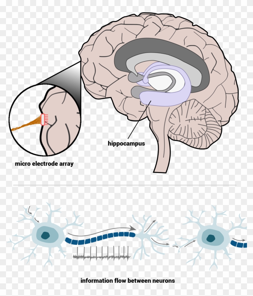
Cross Section Illustration Of Human Brain Showing Limbic ...
Anatomy of the Brain Chart 20x26. The 20" x 26" (51 x 66 cm) chart beautifully illustrates cerebral hemispheres, lobes of the brain, cross section of meninges and venous sinuses, typical nerve cell, synapses, typical glial cells, arteries of the brain, vagus nerves, circulation of cerebrospinal fluid, midbrain with corpus callosum, sagittal ...
Cross Section Of The Human Brain, HD Png Download - 1183x1328 ...

Human skull cross-section with brain, illustration - Stock ...
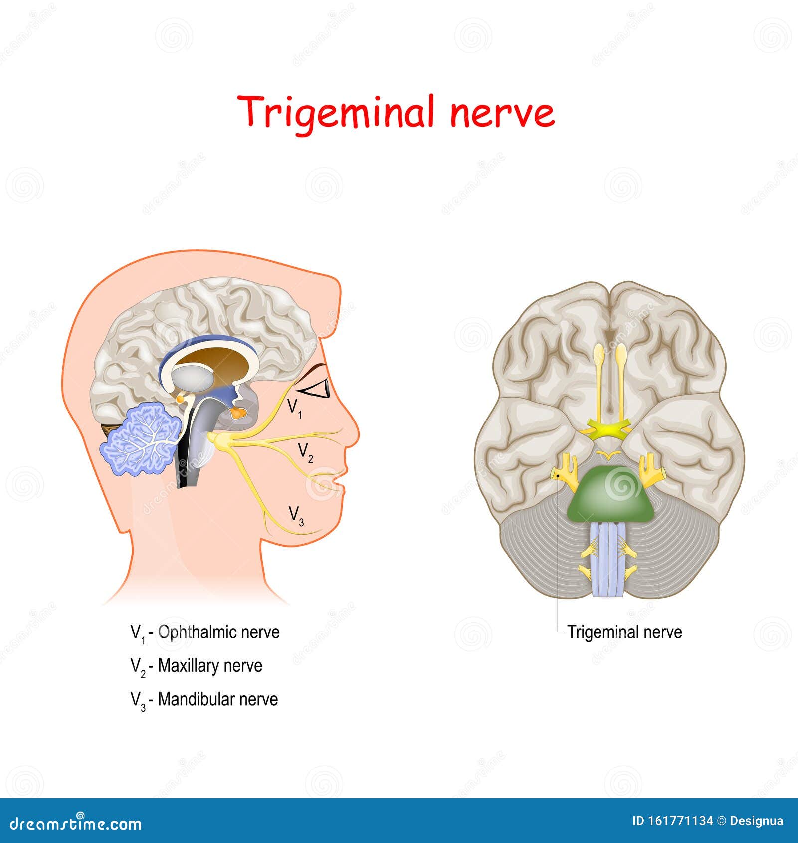
Trigeminal Nerve. Head Cross Section and Bottom View of the ...
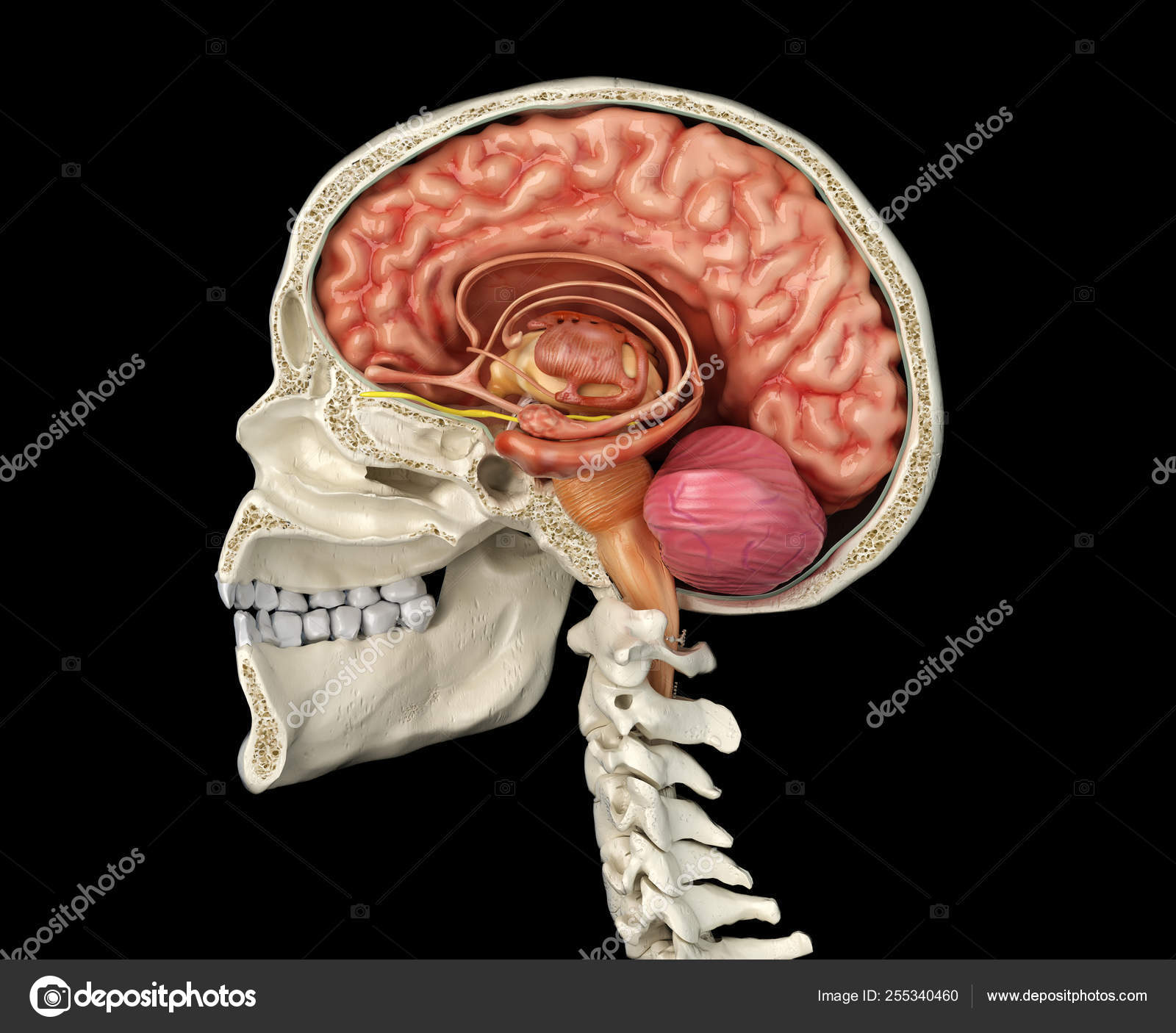
Human skull cross section with brain. Stock Photo by ...

387 Brain Cross Section Diagram Pictures Stock Photos ...

Amazon.com: Functional Human Brain Model, Cross Section - 1/2 ...

The blood-brain barrier-diagram of a cross-section of the ...
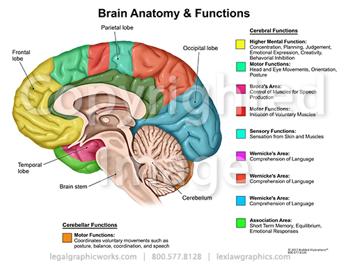
Brain Cross Sectional Anatomy & Functions - Legal Graphicworks
Brain anatomy cross section Images, Stock Photos & Vectors ...
Human brain cross-section — nervous system, central nervous ...
human brain cross section diagram. 3d render, illustration ...

Cross section of human eye, eps8 | Human brain anatomy, Brain ...
Shutterstock - PuzzlePix

Brain Cross section Diagram | Quizlet
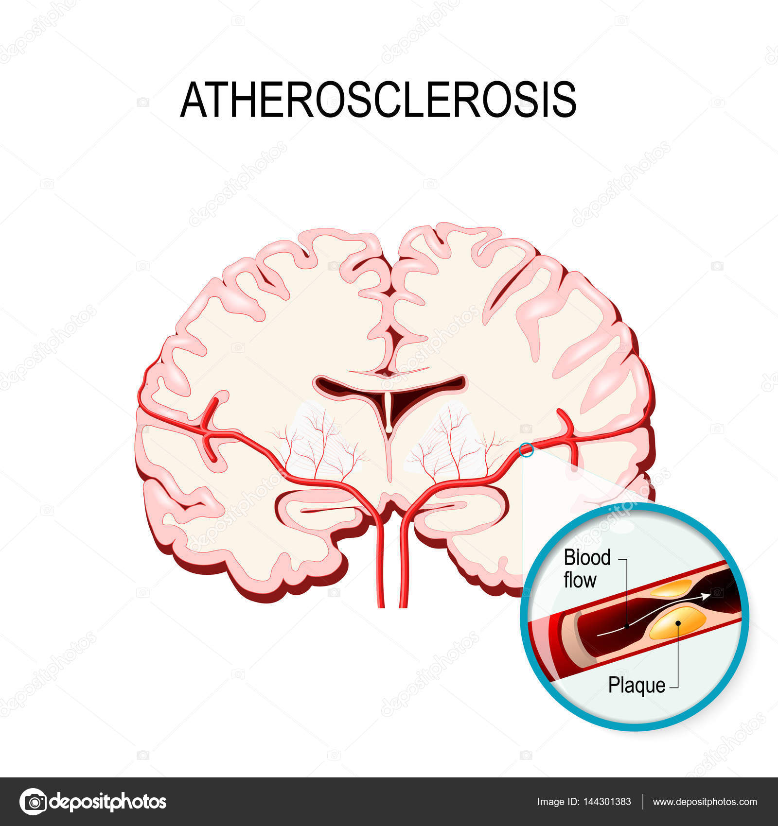
Cross section of the brain Human brain with atherosclerosis ...
Short-Term Memory Loss: Definition, Causes & Tests | Live Science
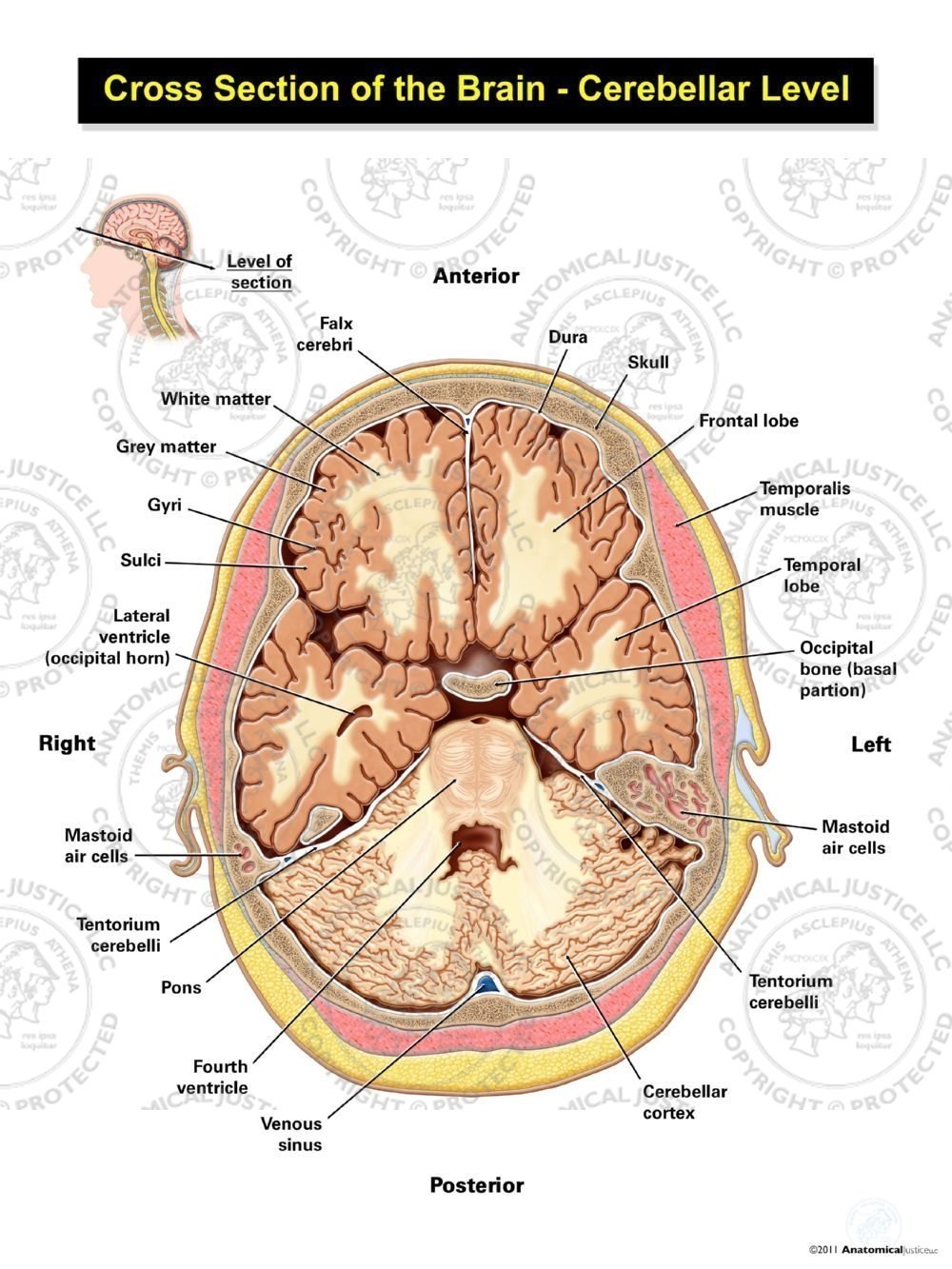
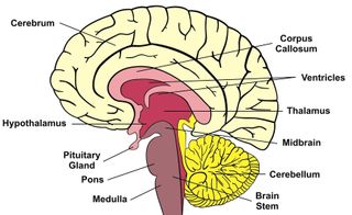
Cross Section of the Brain - Cerebellar Level

Human Brain Anatomy Diagram art print poster
![Brain cross section - Stock Illustration [65838488] - PIXTA](https://en.pimg.jp/065/838/488/1/65838488.jpg)
Brain cross section - Stock Illustration [65838488] - PIXTA

Human brain cross section, illustration Illustration of a ...
brain diagram cross section - Clip Art Library
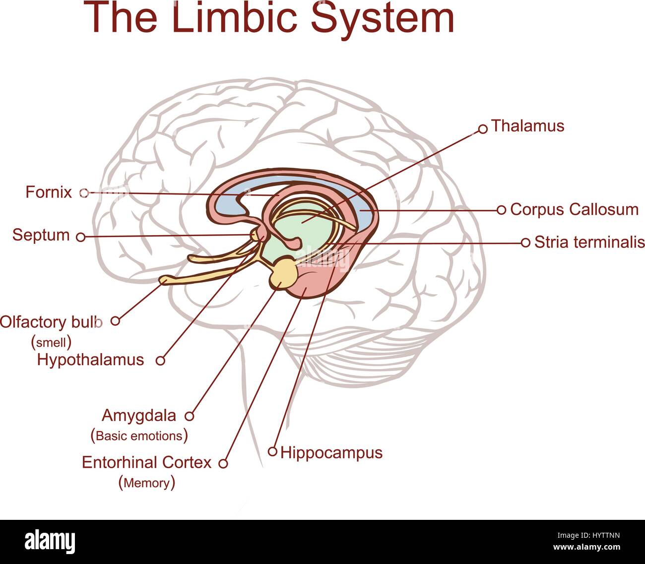
Cross section through the brain showing the limbic system and ...

Human brain cross-section, illustration - Stock Image - F016 ...

Pineal gland anatomical cross section vector illustration ...

cross-section brain diagram | Brain diagram, Human brain ...
5,453 Brain Cross Section Stock Photos, Pictures & Royalty ...

Brain Cross Section Transparent, HD Png Download ...

Cross section illustration of the brain Stock Photos and ...


Comments
Post a Comment