39 diagram of sheep brain
Dr parker s a p i sheep brain dissection youtube lateral view of the with window centered at superior right orbit neuroanatomy diagram cow eye images how to guide and refrence. Sheep Heart Dissection. Sheep have a four-chambered heart, just like humans. By studying the sheep’s anatomy, you can learn how your own heart pumps blood through your body, thereby keeping you alive! Use this sheep heart dissection guide in a lab for high school students. You can also look at the labeled pictures to get an idea of what the ...
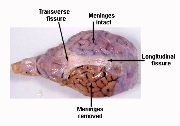
The sheep brain is exposed and each of the structures are labeled and described in a sequential manner, in the same way that a real 1. The sheep brain is enclosed in a tough outer covering called the dura mater. You can still see some structures on the brain before you remove the dura mater.

Diagram of sheep brain
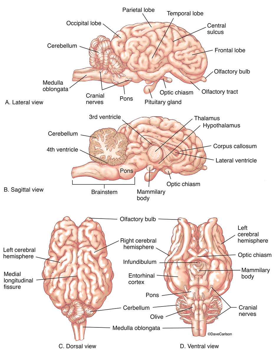
will stimulate a placental and fetal inflammatory response associated with brain injury, which will be exacerbated by chronic hypoxemia and low-grade infection. Chronically instrumented fetal sheep served as controls or underwent repetitive UCOs for up to 4 hours or until fetal arterial pH was <7.00. Normoxic-UCO and hypoxic-UCO fetuses had ... Sheep Brain Explora/on Guide Sheep Brain Exploration Introduction: This guide is intended to lead you through the anatomy of the sheep brain dissection and also to make As illustrated in the human brain diagram below, there are two optic nerves that bring visual information from the eye to the brain. The brain of the sheep is useful for study because its anatomy is similar to human brain anatomy. Although exact proportions (and names) sometimes differ, every structure you will identify in the sheep brain corresponds to a homologous structure, usually with the same name, in humans.
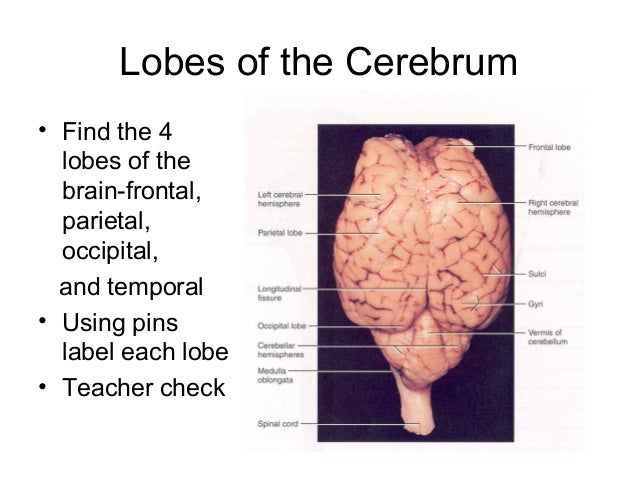
Diagram of sheep brain. An elephants brain is about four times the size of a humans brain. (See diagram on the left). Out of all the animals that have ever lived on earth, the brain of the elephant is the largest known. Elephants are born with 35% of the mass of the adult brain. The elephant is among the more intelligent animals. The brain weight of the male African elephant is 4.2-5.4 kilograms. The … Those students participating in Sheep Brain Dissections will have the opportunity to dissect and compare anatomical structures. At the end of At the end of this document, you will find anatomical diagrams, vocabulary review, and pre/post tests for your students. The following topics will be... Lesson Summary: Dissecting a sheep brain, students gain. appreciation for the complexity of the brain. • engage in scientific observations. • identify the major parts of the brain and explain their functions. • explain what comprise the white and gray matter. • practice dissection and drawing... Labeled Sheep Brain Diagrams Hol Brain Anatomy Nervous System Anatomy Basic Anatomy And Physiology. Image Result For Sheep Brain Labeled Brain Sheep Brain Dissection Project Guide Hst Learning Center Dissection Life Science Lessons Brain Based Learning. Dorsal View Of Sheep...
Human Brain Diagram. Cranial Nerves Coloring. Sheep Brain: Olfactory Tracts (CN I), Optic nerve (CN II), Olfactory Bulbs, Cerebellum, Pons, Medulla. Start studying Sheep Brain. Learn vocabulary, terms and more with flashcards, games and other study tools. the path along which the olfactory receptors send their electrical messages to the brain. Sheep brains have less ridges and contours in comparison to human brains. While a human brain has more of a rounded shape, a sheep's brain is Sheep brains do not have these capabilities. Metacognition and other advanced cognitive skills, such as social intelligence, planning and reasoning... Sheep Brain — Median View. Brainstem components of the ventricular system are visible in this median view of a sheep brain. The third ventricle is indicated by yellow pics.
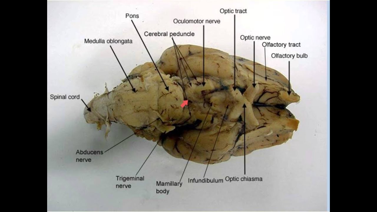
In your own words, describe the firmness and texture of the sheep brain tissue as observed when cutting into IT. Felt tough, and rubbery. When cutting into, IT gets alot softer as you cut through. Sheep brains are used in this lab because they are easy to extract, reasonably inexpensive (they are procured from the food industry), large, and mammalian. A structure is anterior to another structure when it is closer to the nose of an animal (see the above diagram). Some texts use the terms anterior... Sheep Brain Dissection Project Guide - HST Learning Center. 8 hours ago Diagram Worksheets. Label the Parts of a Sheep Brain. Print out these diagrams and fill in the labels to test your knowledge of sheep brain anatomy. Internal anatomy: label the right side (.pdf) External anatomy: label the top... Diagram showing features of cerebral outer membranes and supply of blood vessels. The internal carotid arteries supply oxygenated blood to the front of the brain and the vertebral arteries supply blood to the back of the brain. These two circulations join in the circle of Willis, a ring of connected arteries that lies in the interpeduncular cistern between the midbrain and pons. The …

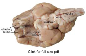
inferior view of sheep brain
Let’s look at the life cycle of Ascaris lumbricoides with a neat and labelled diagram to get a better understanding. Life Cycle: 1) The Egg – Stage 1 – The adult Ascaris worms live inside the walls of the small intestine in human beings. The female worm grows up to 35 cm in length and lays approximately 20,000 eggs that are passed out of ...

sheep brain by Melanie Brawley - Issuu
The sheep brain is quite similar to the human brain except for proportion. Compare the sheep brain to the human brain. What do you notice about the size difference of each structure? Identify the clublike olfactory bulbs on the inferior surface of the frontal lobes of the cerebral hemispheres.

Brain Dissection Guide
Study Flashcards On Lab Exam 3: Anatomy of Sheep Brain; Histology at Cram.com. Quickly memorize the terms, phrases and much more. Diagram of sheep brain, dorsal view. Neuron cells (400x). Nerve and blood vessels slide (closeup of nerve).

Live From Surgery: Sheep Brain Dissection – Join Us Live! | Facebook
sheep Brain Whitney Ewing Melissa Hines Pierra Smith Diagram Human Brain Vs. Sheep Brain Sheep brains do not have as many ridges and contours when compared to human brains, that have a considerable number of ridges and contours to give them an apparently much larger area than the.
Sheep Brain Dissection
Metastasis or dissemination to other organs (e.g., lungs, brain, heart, bone) may occur if protoscolices are released from cysts, sometimes called “secondary echinococcosis.” Neotropical Echinococcosis (Echinococcus vogeli, E. oligarthrus) The Neotropical agents follow the same life cycle although with differences in hosts, morphology, and cyst structure. Adults of E. vogeli …

Image result for sheep brain labeled | Nursing study guide ...
We hope this picture Sheep Brain Sagittal Section Medial View can help you study and research. for more anatomy content please follow us and visit our Seizure Epilepsy Helping Steps Diagram. January 27, 2022Medical skills,Neurology Disease anatomy. Liver Cirrhosis Signs And Symptoms.
Sheep Brain | Carlson Stock Art
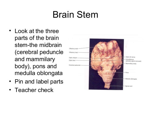
8. Human vs Sheep • Compare the various areas of the sheep brain (cerebrum, brain stem, cerebellum) to the human brain. #4. How is it the same and How is it different? Label your diagram with parts of midbrain and brain stem • What is the function of the medulla oblongata and pons? •

One hemisphere of sheep brain that is cut into four blocks ...
In this video, Natalie demonstrates how to extract a sheep brain. The brain is then used for brain tanning deer hides.Are you interested in learning how to...

Sheep Brain Images
Unraveling the principal brain texture features of preclinical models that are advantageously exploited in experimental neuroscience is crucial to correctly evaluate investigational findings and to correlate them with real clinical scenarios. Although structurally similar to the human brain, the gyrencephalic ovine...

Sheep Brain Diagram
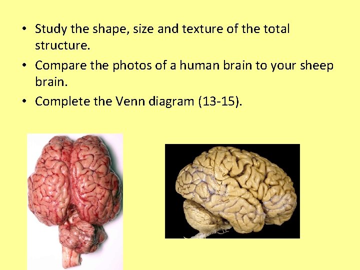
Human vs Sheep Brain There are a few differences between the human and sheep brain. The human brain is larger in size and shape when compared to the sheep's brain. Sheep brains do not have as many...
BRAIN ANATOMY: 101 SHEEP BRAIN DISSECTION!
22.08.2021 · In the labeled diagram, I showed you the skull bone, vertebrae (cervical to caudal), ribs, sternum, wing bones, and leg bones from a chicken. You may also get help from the video that I will add at the end of this article. That video might help you to identify all the bones of a chicken. Chicken skeleton anatomy diagram. Chicken bone anatomy. In each bone from …
49 Sheep Brain Dissection.pptx
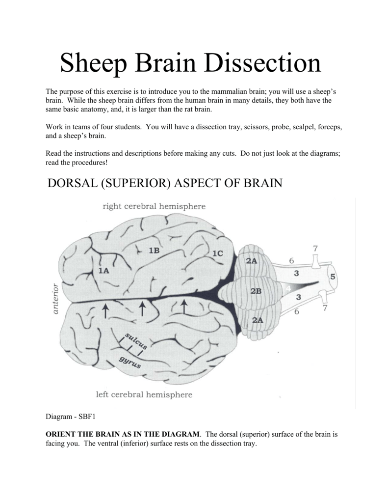
Sheep brain dissection: pre-lab. (Pre-Lab must be submitted to start the lab). Part 1: Planes and Axis of the Brain. 1. Label the diagram to the right with the dorsal-ventral axis and the anterior-posterior axis. 2. Name the view of the brain shown in the diagrams below

Sheep Brain Images
Nervous system - sheep brain images. Sheep Brain Unlabeled.
Sheep+brain+dissection
Compare the size of the olfactory bulbs in a sheep relative to the total brain size compared to the Think about the different components of a neuron (see diagram below) and how this may influence Observe the sheep brain that has had the cerebellum and the caudal portion of the cerebral cortex...
Sheep Brain Dissection
Menstrual cycle is a part of reproductive events in primate females. Know here all about Menstrual cycle definition, phases, reasons, duration, diagram, etc.
Lab: Sheep Brain Dissection
Sheep Brain Dissection Project Guide | HST Learning Center. Learn the external and internal anatomy of sheep brains with HST's Learning Center science lesson and guide! Diagram worksheets also included.
Sheep Brain Dissection Project Guide | HST Learning Center
12.11.2019 · Add all the sides of the diagram and the answer will be “58” 10+10+15+15+4+4 = 58: BRAIN OUT Level 83 [CREATE A RECTANGLE] Click on the square and move it out to half of the screen. BRAIN OUT Level 84 [A simple question! MAKE THE FOLLOWING EQUATION HOLD] Just click on the right “3” then another 3 will pop out.
11.7: Sheep Brain Dissection - Biology LibreTexts
Sheep Brain. Sheep Brain (1:49). Created by: Cecilia Yu '07 Maintained by: CarolAnn Paul Date Created: July 1, 2004 Last Modified: August 25, 2004 Page Expires: August 6, 2006.
sheep brain anatomy
The purpose of the sheep brain dissection is to familiarize you with the three-dimensional structure of the brain and teach you one of the great methods of studying the Sheep Brain Dissection Guide. Planes of Orientation In addition to the direction, the brain as a three dimensional object can be divided.
Sheep Brain Dissection with Labeled Images
Test your brain skills with slide puzzle games. Instead of collecting and finding traditional puzzle pieces, test your skills with the difficult format of puzzle gaming. The puzzle is fixed in a frame and only one piece can be moved at a time. And when you move a piece, it also changes the position of the others, which makes it more challenging ...

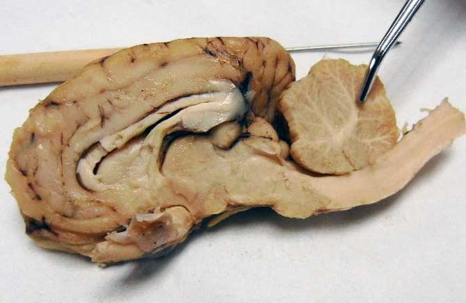
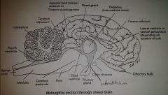
Sheep Brain Midsagittal Section
The brain of the sheep is useful for study because its anatomy is similar to human brain anatomy. Although exact proportions (and names) sometimes differ, every structure you will identify in the sheep brain corresponds to a homologous structure, usually with the same name, in humans.

Sheep Brain Dissection Review Internal Structures. - ppt download
Sheep Brain Explora/on Guide Sheep Brain Exploration Introduction: This guide is intended to lead you through the anatomy of the sheep brain dissection and also to make As illustrated in the human brain diagram below, there are two optic nerves that bring visual information from the eye to the brain.

Sheep Brain Dissection 1 Diagram | Quizlet
will stimulate a placental and fetal inflammatory response associated with brain injury, which will be exacerbated by chronic hypoxemia and low-grade infection. Chronically instrumented fetal sheep served as controls or underwent repetitive UCOs for up to 4 hours or until fetal arterial pH was <7.00. Normoxic-UCO and hypoxic-UCO fetuses had ...
Brain Dissection LAB How to perform a successful
Sheep Brain Dissection Lab Companion
Medical Detectives Lesson 28

Sheep Brain Images

Sheep brain images | Lab | Amherst College

Lateral Sheep Brain Diagram | Quizlet
Sheep+brain+dissection
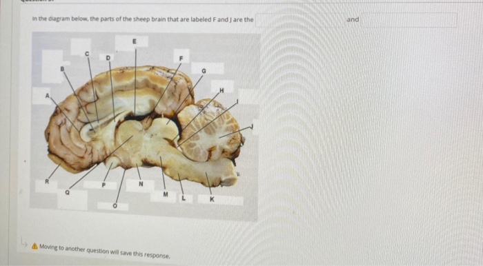
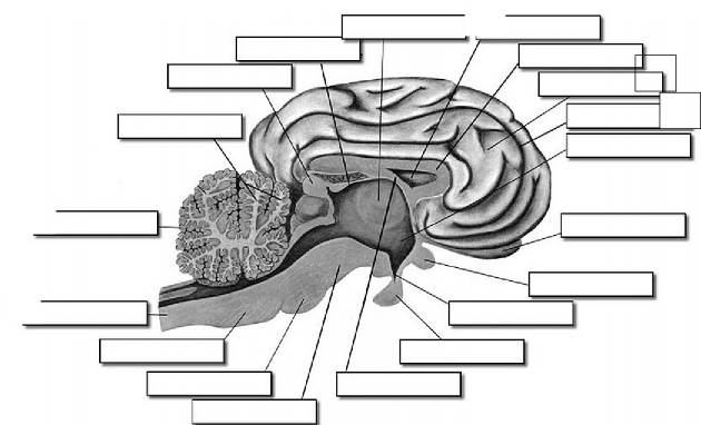
Solved in the diagram below, the parts of the sheep brain ...
Lab: Sheep Brain Dissection
Human Brain Sheep Frontal Lobe Lobes Of The Brain PNG ...

Cerebrum Sheep Dissection - Human Anatomy - GUWS Medical

Sheep Brain Dissection - DocsBay

Image result for sheep brain labeled | Brain diagram, Human ...

Sheep Brain Dissection

Superior View Of Sheep Brain - Sheep Brain Superior View ...
Lab Exam 3: Anatomy of Sheep Brain; Histology Flashcards ...
Comments
Post a Comment