39 eeg lead placement diagram
3 lead ECG cable Placement (there are two ways) Way 1. Monitors one of the three leads: RA: placed the red electrode within the frame of rib cage,right under the clavicle near shoulder( see chart in follow picture) LA: the yellow electrode is placed below left clavicle, which is in the same level of the Red electrode • Describe specific measures to make a child more comfortable in the EEG lab, in preparation for head measurement and lead placement • Explain lead placement techniques to ensure good lead placement without injury to the child • List the pros and cons of using sedation for lead placement for the pediatric patient,
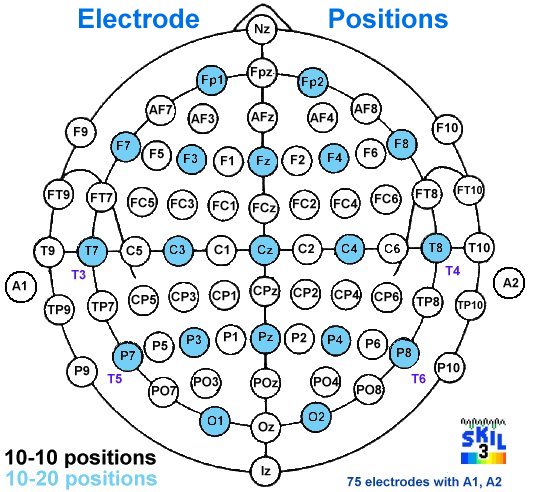
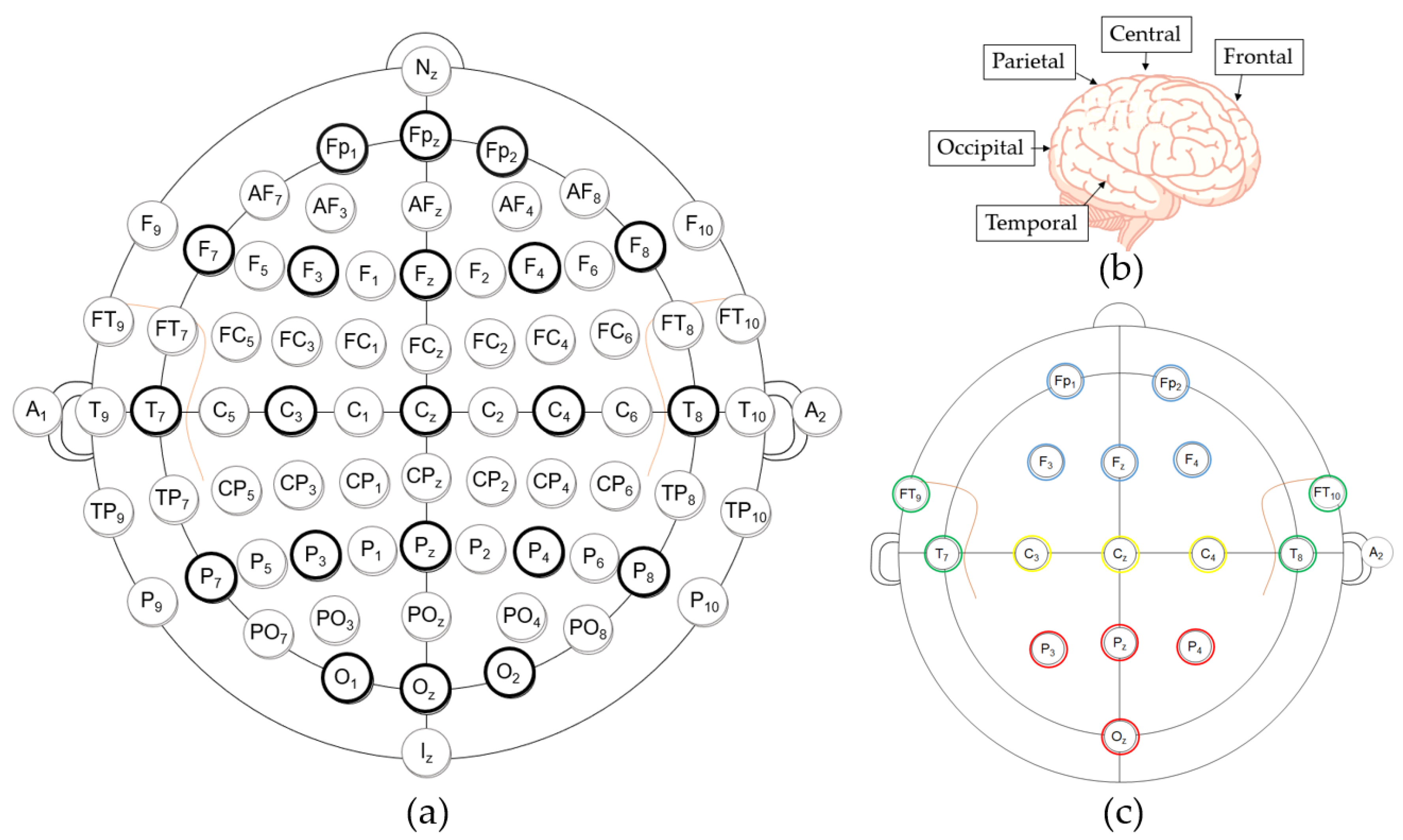
With the availability of EEG systems capable of recording with a greater number of channels (e.g. 128, 256), there is a need to standardize placement of additional electrodes. A further extension of the 10-10 system, called the 10-5 system, has been proposed, 4 but

Eeg lead placement diagram
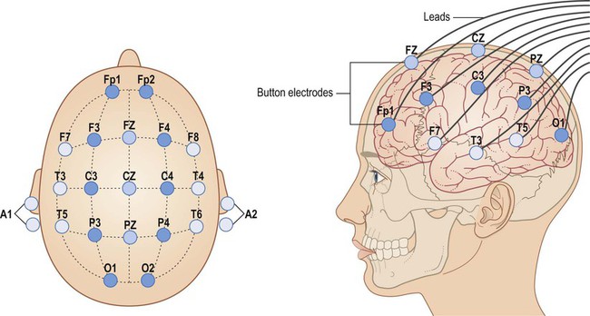
eeg electrode placement system: The most popular scheme used in the placement of electrode for the EEG pick up is the 10-20 electrode placement system. As the cranial area is divided into four main regions, the electrodes are placed accordingly in the different regions. Additional notes on 12-lead ECG Placement: The limb leads can also be placed on the upper arms and thighs. However, there should be uniformity in your placement. For instance, do not attach an electrode on the right wrist and one on the left upper arm. For female patients, place leads V3-V6 under the left breast. Place one sub-hairline electrode in the centre of the sub-hairline 2. Place one electrode in front of each ear 3. Place one electrode over each temple 4. Place one electrode between the centre electrode and the temple electrode on each side 5. Place one electrode behind each ear Electrode Placement Identify the CEEG Lead Colors FP2 Ref Right T6
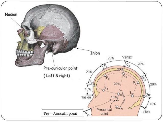
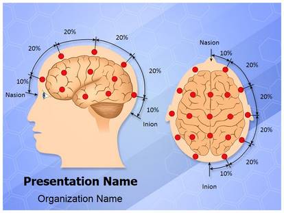
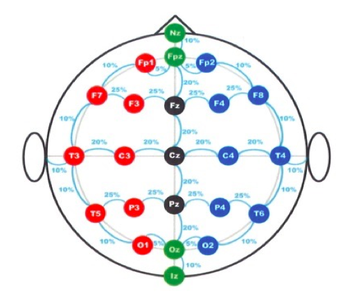
Eeg lead placement diagram. Placement of paediatric ECG leads In young children, the right ventricle normally extends to the right side of the sternum. To appropriately display right ventricular potentials, ECGs for children in the under five-year age group must include an alternate lead ( 'V4R') on the right side of the chest, at a point analogous to the left-sided V4. Placement of Lead V1 Locate the sternal notch (Angle of Louis) by feeling the top portion of the breast bone, and moving your fingers downward until you feel a bump. Move your fingers to the right, off of the bump, and you will feel some soft tissue in between the 2nd and 3rd rib. This is the 2nd intercostal space. EEG Lead System. International Federation of EEG society has suggested 10 - 20 electrode placement system for EEG recording.Silver / silver chloride electrodes are used as surface electrodes in this setup. On the scalp, distances between two electrodes are given as 10% and 20% of the distance between specified points. 1 - 6 A third electrode placement, termed bifrontal, in which the electrodes are placed on either side of the forehead above the eyes, has come into use but there are no data about any possible neurophysiological differences as reflected in the ictal EEG with this placement vis a vis the other two placements.
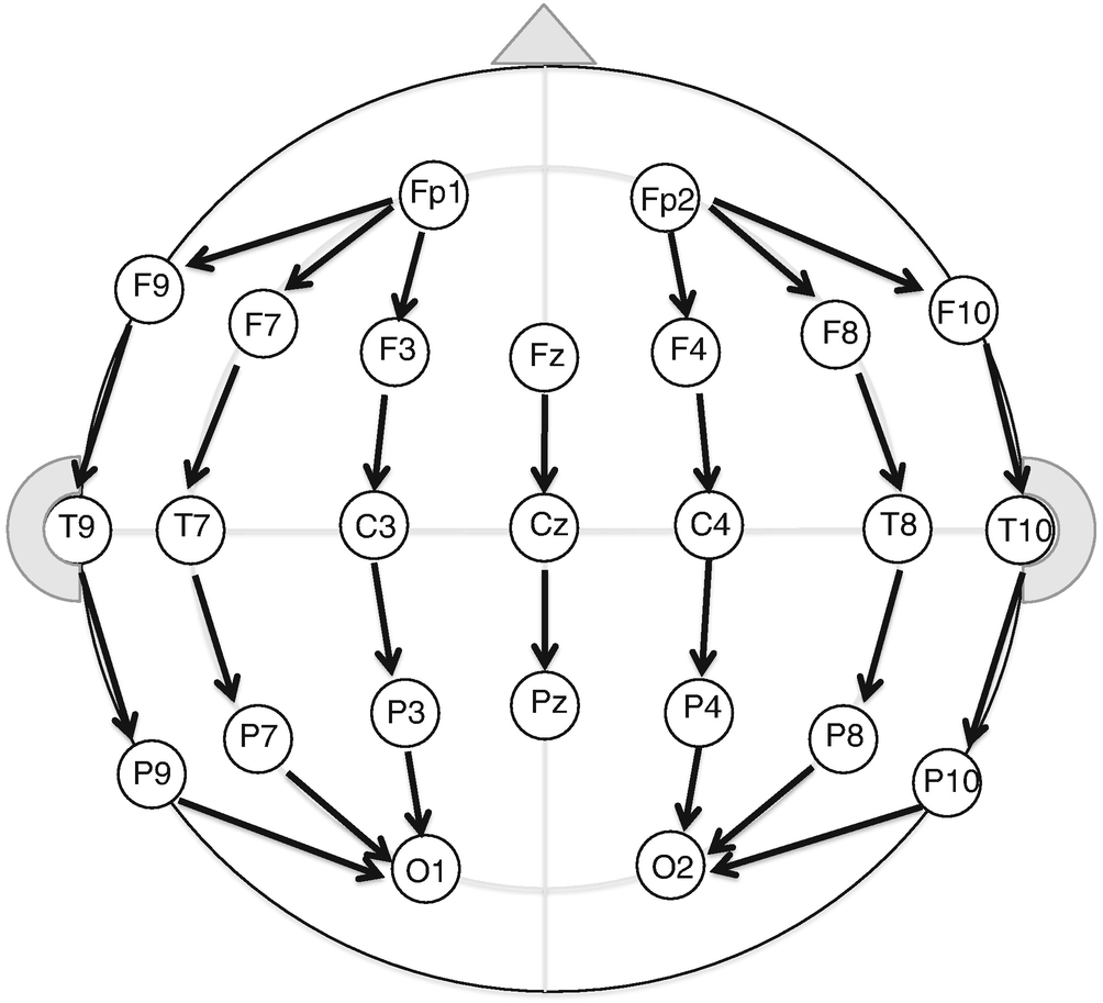
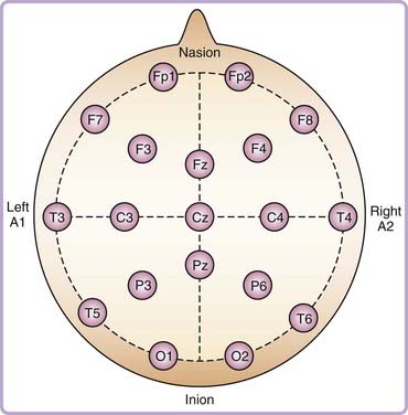
The first step to any EEG study is the placement of the electrodes, and this is most commonly done via the international, standardized 10-20 System. This system is so named because it splits the skull into increments of 10% or 20% to place the electrodes, ensuring that each electrode is relatively positioned to all the others and making it ... • EEG laboratory is a new experience for most individuals -Assume participant knows nothing of EEG/MEG -Give enough information in everyday language -Explain the length of each experiment phase -Leave time for questions • Working with children -Criminal records check (show it to Anna Karhu, Jutta Aalto or Raija Mehto) positioning of the electrodes: first, the nasion which is the point between the forehead and the ... Even numbers (2,4,6,8) refer to electrode positions.20 pages EEG has very high time resolution and captures cognitive processes in the time frame in which cognition occurs. ... Electrode arrays and placement. ... they take the lead and apply automated de-noising procedures or automatically generate high-level cognitive-affective metrics which can be used in order to get to conclusions much faster.
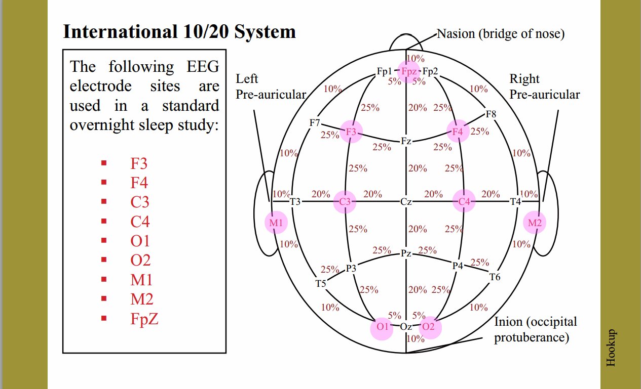
Using 3 referential EEG derivations during PSG, as recommended in the AASM manual, instead of a single central EEG derivation, as originally suggested by Rechtschaffen and Kales (1968), resulted in a mean ± SE decrease in N1 sleep of 9.6 ± 3.9 min (P = 0.018) and an increase in N3 sleep of 10.6 ± 2.8 min (P = 0.001). The International 10-20 system of electroencephalogram (EEG) lead placement. In neonates, only the shaded electrodes are placed on the smaller neonatal scalp for conventional EEG. Fp3 Fp4 was used... EEG electrode placement is a critical part of successful qEEG. Traditional EEG lead placements follow the 10-20 system, an internationally recognized standard for applying the electrodes attached to your scalp. "10-20" refers to the distance between EEG leads being 10% or 20% of the total distance of the skull. The 10-20 system or International 10-20 system is an internationally recognized method to describe and apply the location of scalp electrodes in the context of an EEG exam, polysomnograph sleep study, or voluntary lab research.This method was developed to maintain standardized testing methods ensuring that a subject's study outcomes (clinical or research) could be compiled, reproduced, and ...
The 10-20 International system of EEG electrode placement ...
Place the electrodes. The lead wires should be attached before the electrodes are applied. The electrodes should be applied by pressing around the edges instead of in the centre (this spreads the gel out and creates air pockets). Correct placement of electrodes is crucial to ensure that the information gathered is accurate.

The standardized EEG electrode array of the IFCN - ScienceDirect
by A Morley · 2016 · Cited by 16 — What is the 10-20 system? • An internationally recognised method that allows EEG electrode placement to be standardised. • Ensures inter-electrode spacing is ...34 pages
Electrode Placement Systems and Montages | SpringerLink
12-Lead ECG Electrode Placement Explained. One of the most common questions related to 12-lead ECG electrode placement is why there are only 10 electrodes. It's very important to understand what the term "lead" really means. A lead is a view of electrical activity of the heart from a specific angle across the body.

Electrodes placement according to the 10-20 International ...
10-20 system EEG Placement Andrew Morley (BSc Hons, RPSGT)(BSc Hons, RPSGT) , Lizzie Hill Lizzie Hill (EST RPSGT) & Prof. Dr Athanasios G. Kaditis. Chief Respiratory (Sleep) Physiologist, Royal Hospital for Children, Glasgow. Specialist Respiratory Clinical Physiologist, Royal Hospital for Sick Children, Edinburgh
EEG electrode placement (red circles). The diagram is a ...
Electroencephalography (EEG) is a method to record an electrogram of the electrical activity on the scalp that has been shown to represent the macroscopic activity of the surface layer of the brain underneath. It is typically non-invasive, with the electrodes placed along the scalp. Electrocorticography, involving invasive electrodes, is sometimes called intracranial EEG.
10-20 electrode placement system, EEG - video Dailymotion
EEG disk electrodes, however, require the use of a conductive cream. Within a few days of application, the cream will dry or be absorbed, causing higher im-pedances. This process requires re-prepping and/or re-application of electrodes.
10-20 EEG Placement
Press 1 lead onto the left shoulder below the clavicle. As on the right arm, this lead should fall about 2 inches (5.1 cm) below the clavicle. Place the lead at roughly the same location as you placed the lead on their right side to ensure that both arm leads give accurate readings. Press down firmly on the lead to fix it in place.
Epilepsy | Neupsy Key
This section describes placement of a frontal electrode Fz, a central electrode C4, and an occipital electrode O1. Backup electrodes at F4, C3, Cz, and O2 are recommended in case an electrode falls off. Signals from the EEG electrodes are referred to an electrode placed over the contralateral mastoid.
Introduction to the electroencephalogram (EEG) (Chapter 14 ...
Electrodes: Gold cup are typically used for EEG experiments Electrode Placement: Typically, EEG electrodes are placed based on the 10-20 method. The 10 and 20 in the name refer to the percent distances that the electrodes are from each other in proportion to the size of the head.

A Novel Method for Stress Measuring Using EEG Signals ...
•Primer of EEG with a Mini-Atlas. AJ Rowan and E Tolusky. Butterworth Heinemann 2003 •Atlas of EEG in Critical Care. LJ Hirsch and RP Brenner. Wiley Blackwell 2010 •Prognostic value of continuous EEG monitoring during therapeutic hypothermia after cardiac arrest. AO Rossetti et al. Critical Care 2010 (14): R173

10–20 system (EEG) - Wikipedia
recording of sleep requires measurement of brain activity (EEG), eye mov ement (EOG) and muscle activity (EMG). On the following page are examples of how the following stages appear on a polygraph record of EEG: Wakefulness - (Awake and Drowsy patterns) Note how irregular the pattern looks.
10 and 20 electrode placement
Electrode Placement. Accurate placement of many electrodes on the scalp is time consuming and requires practice. EEG caps greatly facilitate this process. These caps are made of elastic fabric (often available in different sizes), and electrodes are already fixed in the proper configuration.
Free Eeg Electrode Placement Medical PowerPoint Template for ...
EEG Electrode Placement. Create healthcare diagrams like this example called EEG Electrode Placement in minutes with SmartDraw. SmartDraw includes 1000s of professional healthcare and anatomy chart templates that you can modify and make your own. 8/37 EXAMPLES. EDIT THIS EXAMPLE. CLICK TO EDIT THIS EXAMPLE.

10-20 Electrode Placement Map | Highlighted Regions | Brain ...
Place one sub-hairline electrode in the centre of the sub-hairline 2. Place one electrode in front of each ear 3. Place one electrode over each temple 4. Place one electrode between the centre electrode and the temple electrode on each side 5. Place one electrode behind each ear Electrode Placement Identify the CEEG Lead Colors FP2 Ref Right T6

Eeg Electrode Placement PowerPoint Presentation Template With ...
Additional notes on 12-lead ECG Placement: The limb leads can also be placed on the upper arms and thighs. However, there should be uniformity in your placement. For instance, do not attach an electrode on the right wrist and one on the left upper arm. For female patients, place leads V3-V6 under the left breast.
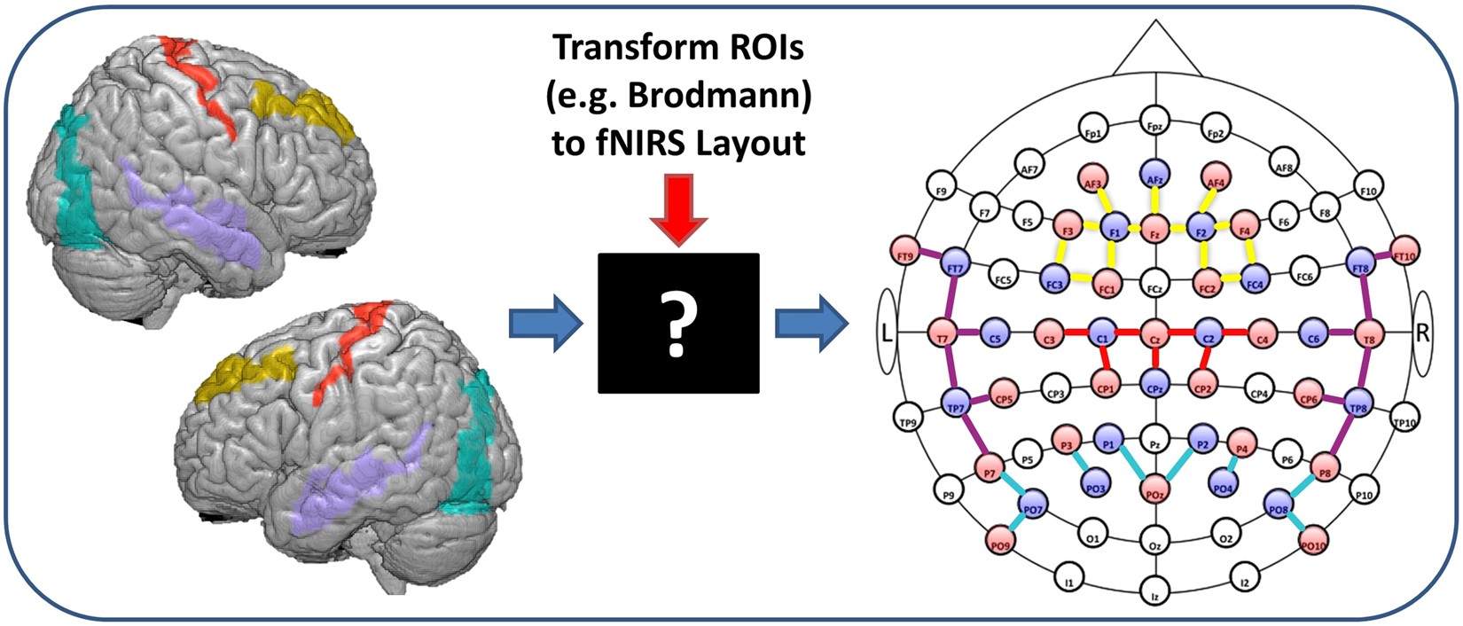
fNIRS Optodes' Location Decider (fOLD): a toolbox for probe ...
eeg electrode placement system: The most popular scheme used in the placement of electrode for the EEG pick up is the 10-20 electrode placement system. As the cranial area is divided into four main regions, the electrodes are placed accordingly in the different regions.
ECG/ EKG
EEG: electrode positions & Broadmann atlas - minks - åšå®¢å›

Electrode positions | Details | Hackaday.io

EEG electrode placement and the 4 ROIs. | Download Scientific ...

Using a structured-light 3D scanner to improve EEG source ...

EEG electrode placement, eps10 canvas print

EEG Electrode Placement: Fixed vs. Variable | Bitbrain

EEG electrode placement - UpToDate
Sensors | Free Full-Text | Drowsiness Detection Based on ...

Figure 19. [The 10-20 System electrode placements ...

Choosing your reference for an EEG recording and the ...

Understanding EEG Part 4, 10-20 electrode placement system used in electroencephalography (EEG) test

EEG electrode placement | Sleep lab, Sleep medicine, Child ...
Neurologic Monitoring Techniques | Neupsy Key
Cost-Effective EEG Signal Acquisition and Recording System
EEG
EEGGuideline 2 Electrode nomenclature American Clinical ...

Cureus | Stereotactic Bony Trajectory Preservation for ...

Study of Resting-State Functional Connectivity Networks Using ...

DWT-Net: Seizure Detection System with Structured EEG Montage ...
Multi-Channel EEG (BCI) Devices
The International 10/20 System of Electrode Placement ...

EEG electrode placement


Comments
Post a Comment