39 liver histology diagram
Apr 15, 2020 - Explore Summer Ekelund's board "Histology - Liver", followed by 312 people on Pinterest. See more ideas about liver, histology slides, liver anatomy. Download scientific diagram | Liver histology (H&E) (A) and immunohistochemistry for CD-45 (B). LPS induced liver perivenulitis. Treatment with simvastatin, especially when given prior to LPS ...
How to draw Liver, stomach, small and large intestine, and cloaca in Biology practical copy.
Liver histology diagram
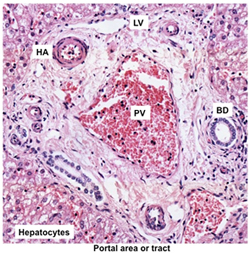
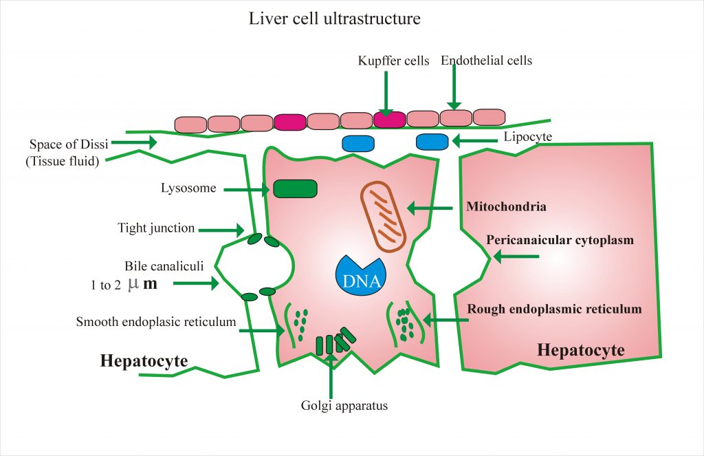
The liver consists of the following major histological components: Parenchyma, which is represented by hepatocytes Stroma, which is a continuation of the surrounding capsule of Glisson. It consists of connective tissue and contains the vessels. The capsule is also covered by a layer of mesothelium, arising from the peritoneum covering the liver. Gallbladder histology. Gallbladder is a hollow, pear shaped organs of digestive system of body. It attached to the lower surface of the liver. You should read this full article to know about the liver histology.This is the ultimate guide to learn normal liver histology in details.. Before to start I would like to provide the summary of this organ. Liver and intrahepatic bile ducts - nontumor - Normal histology. The liver is the largest solid organ and is located in the right upper quadrant of the abdomen. Menu ... Diagrams / tables. Images hosted on other servers: Lobule and portal triad. Anatomy and functional segments.
Liver histology diagram. Slide 194 liver, gall bladder H&E View Virtual Slide Slide 195M liver, gall bladder Masson View Virtual Slide. Upon gross examination of slides 194 and 195M (i.e. with the naked eye or at the lowest power on the virtual microscope) you will see a portion of the gall bladder wall nestled in an indentation of the liver tissue. Examine the wall of ... The liver is a triangular, bilobed structure consisting of a larger right lobe and a smaller left lobe. The falciform ligament separates the two lobes. A layer of fibrous tissue called Glisson's capsule covers the liver. This capsule is covered by peritoneum. This protects the liver from physical damage. It has two main sources of blood: Science. Biology. Biology questions and answers. Introduction. Cirrhosis is defined as the histological development of regenerative nodules surrounded by fibrous bands in response to chronic liver injury, that leads to portal hypertension and end stage liver disease. Recent advances in the understanding of the natural history and pathophysiology of cirrhosis, and in treatment of its complications, resulting in improved management, quality of ...
Liver Labelled Diagram - Liver, Gallbladder, Pancreas and Bile Passage The liver has structural characteristics that are not found in any other internal organ of the human body. One abnormal characteristic is the liver's regenerative abilities. Pieces of the liver can be cut off and it will regenerate new hepatic tissue almost like a lizard's tail! Mouse liver lobes Source: Figures 2 and 3 in Harada, T., et al. "Liver and Gallbladder." Chapter 7 in Pathology of the Mouse. Edited by Robert Maronpot. Vienna, IL: Cache River Press, 1999. ISBN: 188989902X. Images removed for copyright reasons. Start studying Liver histology diagram. Learn vocabulary, terms, and more with flashcards, games, and other study tools. 1. LIVER Normal Histology Rifat Mannan,MD Mount Sinai St.-Luke's Roosevelt Hospital Center, New York. 2. Anatomy • Second-largest organ of the body and the largest gland, weighing about 1-1.5 kg. • Comprises 2% of body weight.
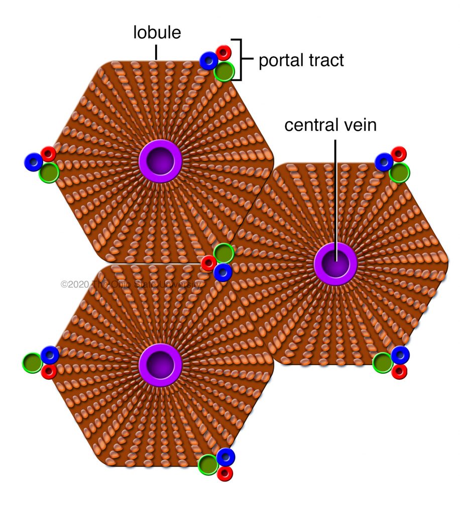
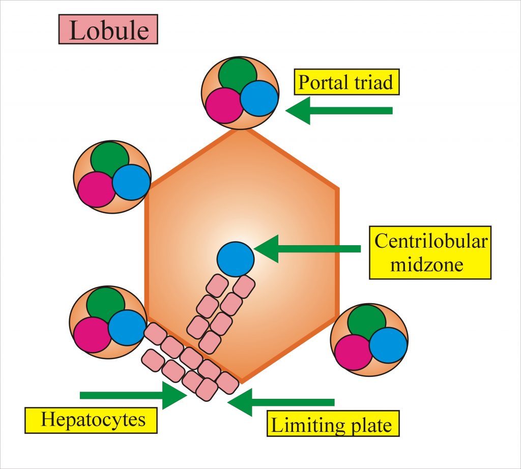
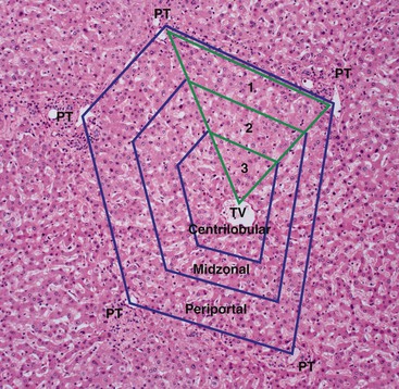
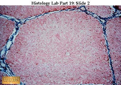
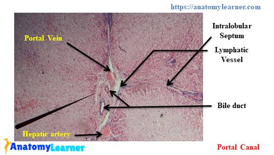
Hepatic Histology: The Lobule. The liver is bounded by a connective tissue capsule which extends into its substance as highly branched septae. The afferent blood vessels and lymphatics follow this connective tissue highway throughout the liver. Efferent vessels traverse a route separate from connective tissue scaffolding. Liver Histology Create healthcare diagrams like this example called Liver Histology in minutes with SmartDraw. SmartDraw includes 1000s of professional healthcare and anatomy chart templates that you can modify and make your own. 46/71 EXAMPLES EDIT THIS EXAMPLE Text in this Example: Liver Histology Three dimensional view of one of the hepatic Our aim was to evaluate whether Voronoi diagrams can be used to describe the classic liver lobular architecture. We examined the histology of normal porcine and human livers and analyzed the geometric relationships of various microanatomic structures utilizing digital tools. Histology of the Liver Looking at a section of liver is somewhat reminiscent of looking down out of an airplane at a suburban neighborhood. One sees a very regular, almost monotonous, collection of houses in blocks demarcated by roads, with a gas station or minimart apparent at almost every intersection.
Structural organization of the liver – Veterinary Histology
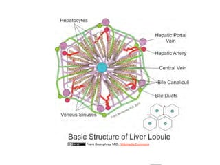
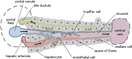
This diagram shows how the relationships between the hepatocytes and the liver sinusoids.. The endothelial cells of the liver sinusoids (capillaries) have pores, including large pores called fenestra.Furthermore the basement membrane is discontinuous and non-obstructing. This allows blood plasma to enter a space between the endothelial cells and the hepatocytes, called the space of Disse ...

Liver Histology Diagram | Quizlet
Liver histology diagram. 6 terms. christinenguyen04. Other sets by this creator. ... Start studying Liver. Learn vocabulary, terms, and more with flashcards, games ...
Liver histology - Labpedia.net
Metabolic toxins are normal by-products of your metabolism, and its the job of the kidneys, liver and lungs to remove these toxins from the blood; therefore ...
Liver Tissue Processing and Normal Histology | Clinical Gate
Chapter 35 Liver & Gallbladder Congenital Conditions GILBERT'S SYNDROME osms.it/gilberts-syndrome PATHOLOGY & CAUSES SIGNS & SYMPTOMS Benign, inherited metabolic disorder; recurring unconjugated hyperbilirubinemia, jaundice Autosomal recessive inheritance pattern AKA Meulengracht disease, familial nonhemolytic jaundice Serum bilirubin increases during physiologic stress (e.g. illness ...

Histology images of Liver & bile ducts by PathPedia.com ...
Download scientific diagram | Histology of normal liver, fibrosis and cirrhosis. A: Representative histological images (using Sirius red staining), normal liver; B: Mild to moderate fibrosis with ...

liver, gall bladder and pancreas
Full labeled anatomical diagrams - Anatomy of the abdomen and digestive system: these general diagrams show the digestive system, with the major human anatomical structures labeled (mouth, tongue, oral cavity, teeth, buccal glands, throat, pharynx, oesophagus, stomach, small intestine, large intestine, liver, gall bladder and pancreas).
01.12.09(b): Histology - Liver, Pancreas, and Gallbladder
Feb 12, 2020 · Hope, you got better idea and ultimate guide to learn the liver histology along with best liver histology diagram and liver histology pdf guide. Now, you may practices and evaluate your learning with the help of liver histology ppt. Here, you will find the liver histology labeled images and other slides for your practices.

liver histology diagram. gilson... - Biology knowledge | Facebook
Liver is the largest organ in the human body, which has several specific functions: metabolic, synthetic, storage, catabolic, and excretory. It is composed of three major compartments, blended together harmoniously: the hepatocytes, the biliary system, and the vascular system. Embryology, gross anatomy, and normal histology of the liver are ...
Histology Slides 1
Histology at Southern Illinois University School of Medicine. Noe, M, Kunz, G, Herbertz, M, Mall, G, Leyendecker, G. The cyclic pattern of the immunocytochemical expression of oestrogen and progesterone receptors in human myometrial and endometrial layers: characterization of the endometrial-subendometrial unit Oxford Journals Human ...

A: Schematic of a typical liver lobule. Adapted under CC BY ...
The liver is a large essential organ found in the upper right quadrant of the abdomen.It is a multifunctional accessory to the gastrointestinal tract and performs such duties as detoxification, protein synthesis, biochemical production and nutrient storage to name but a few. It is the largest gland in the human body, weighing in at approximately 1.5 kilograms.

A: Picture of the anatomy of mouse liver; B-O: Histological ...
Slide 194 liver, gall bladder H&E View Virtual Slide Slide 195M liver, gall bladder Masson View Virtual Slide. Upon gross examination of slides 194 and 195M (i.e. with the naked eye or at the lowest power on the virtual microscope) you will see a portion of the gall bladder wall nestled in an indentation of the liver tissue. Examine the wall of ...
Histology at SIU, liver
Liver and intrahepatic bile ducts - nontumor - Normal histology. The liver is the largest solid organ and is located in the right upper quadrant of the abdomen. Menu ... Diagrams / tables. Images hosted on other servers: Lobule and portal triad. Anatomy and functional segments.

Liver Histology 100x Diagram | Quizlet
Gallbladder histology. Gallbladder is a hollow, pear shaped organs of digestive system of body. It attached to the lower surface of the liver. You should read this full article to know about the liver histology.This is the ultimate guide to learn normal liver histology in details.. Before to start I would like to provide the summary of this organ.

Liver Histology. - ppt download
The liver consists of the following major histological components: Parenchyma, which is represented by hepatocytes Stroma, which is a continuation of the surrounding capsule of Glisson. It consists of connective tissue and contains the vessels. The capsule is also covered by a layer of mesothelium, arising from the peritoneum covering the liver.
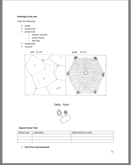
Solved Histology of the Liver Label the following: lobule ...

HISTOLOGY OF LIVER – MANAGE YOUR TIME 1996
Human Structure Virtual Microscopy

Histopathology images of liver. Representative ...

LIver histology | Medical school stuff, Medical laboratory ...

10 Liver ideas | طب, مدرسة, علم

Maternal liver histology. Left column, H&E, 106 magnification ...

Liver Histology Diagram | Quizlet

Mammalian liver histology Diagram | Quizlet
Liver histology - Labpedia.net

Liver: Classic Lobule Model - Histology Flashcards | Draw it ...
Liver - Wikipedia
ZLE ameliorates liver histology of S. mansoni infected CD-1 ...

Liver Organization

Normal appearance of liver histology of control rat ...

Liver Histology
Liver histology: Structure, cells and characteristics | Kenhub

Nutmeg liver. – Histopathology.guru

How to draw histological diagram of Liver in 2 minutes | Rapid Histology | Amit's Lectures | UHS

BIOL 122 Liver Histology Diagram Diagram | Quizlet

Hepatic Pathology
Liver histology - Labpedia.net

5. Representative photomicrographs of liver histopathology ...
Liver Histology – The Ultimate Guide to Learn Liver in Easy ...

Histopathology drawings - Fatty Liver | Facebook


Comments
Post a Comment