43 male pelvic anatomy diagram
Pelvic Bone Diagram and Anatomy This section will extrapolate on the individual anatomy of the bones of the pelvic girdle. The pelvic girdle bones and their location relative to each other can be... Browse 131 pics of the male pelvis diagram stock photos and images available, or start a new search to explore more stock photos and images. Newest results Human Pelvis Image Human male anatomy scheme. Main pelvic bones - sacrum, ilium, coccyx, pubis, ischium. Vector illustration isolated on a white background. Medical and healthcare infographics
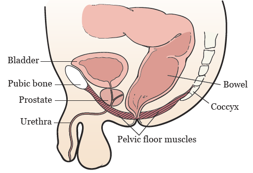
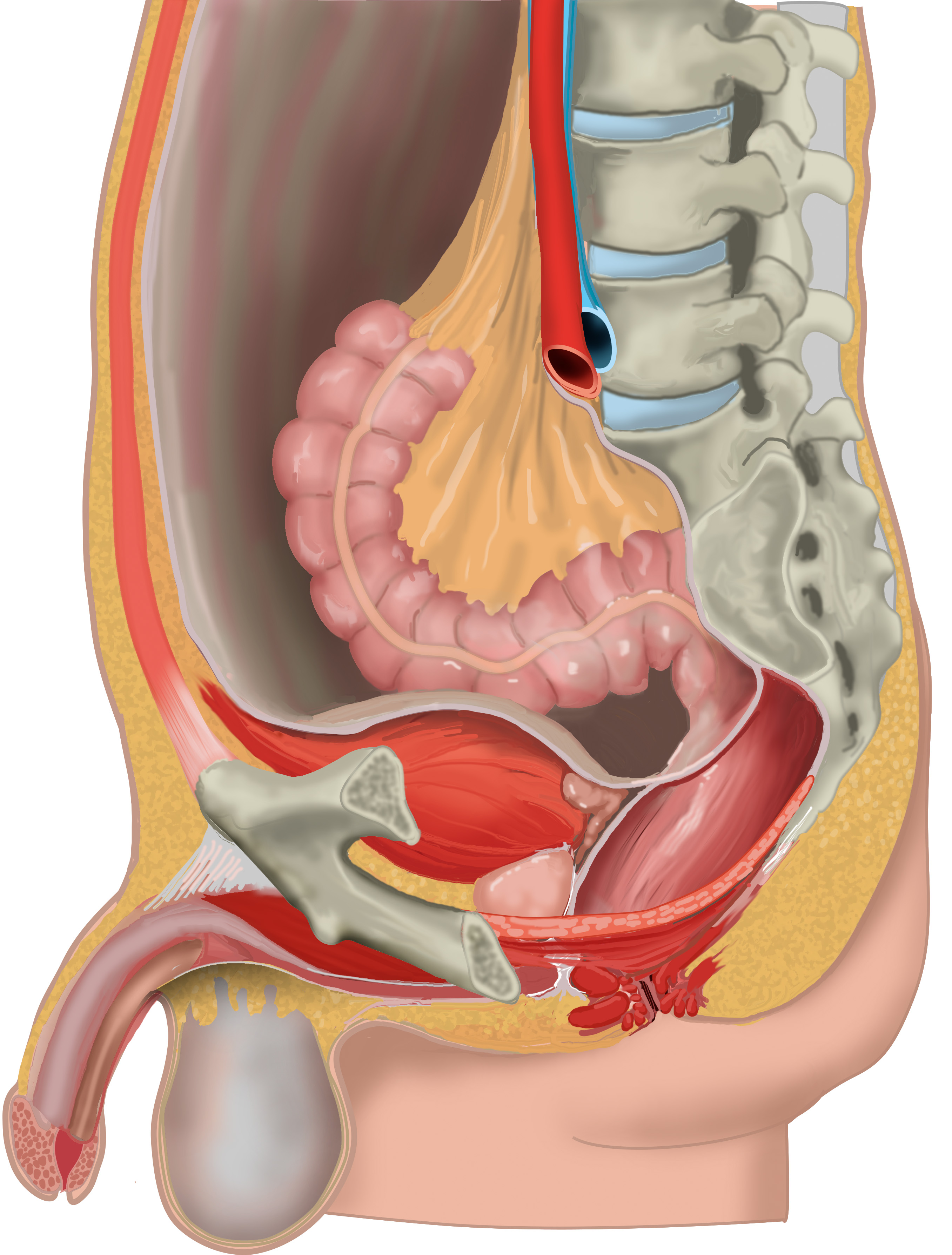
The pelvic diaphragm is a wide but thin muscular layer of tissue that forms the inferior border of the abdominopelvic cavity. Composed of a broad, funnel-shaped sling of fascia and muscle, it extends from the symphysis pubis to the coccyx and from one lateral sidewall to the other.

Male pelvic anatomy diagram
Introduction The male pelvic floor is a complex structure made up of muscles, ligaments, nerves and fascia. The pelvic floor muscles form part of the pelvic floor and play a critical role in sexual function as well as the maintenance of urinary and faecal continence, + This is a course page funded by Physioplus online learning The abdominal viscera occupy the major pelvis; the minor pelvis is the narrower continuation of the major pelvis inferiorly. The inferior pelvic outlet is closed by the pelvic floor. The female pelvis (Figure 1A) has a wider diameter and a more circular shape than that of the male. The wider inlet facilitates head engagement and parturition. The leg of a dog's pelvic limb consists of the tibia and fibula bones. In contrast, the hind paw includes the tarsal bones, metatarsals, and digits that include the three phalanges in each and sesamoid bones. But, how the dog pelvis anatomy is formed? The dog bony pelvis is formed by the ossa coxarum (both sides) and the sacrum bone.
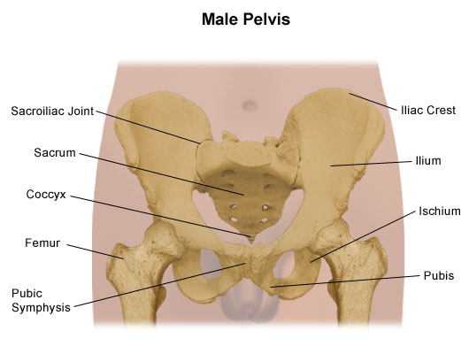
Male pelvic anatomy diagram. pelvis, also called bony pelvis or pelvic girdle, in human anatomy, basin-shaped complex of bones that connects the trunk and the legs, supports and balances the trunk, and contains and supports the intestines, the urinary bladder, and the internal sex organs.The pelvis consists of paired hipbones, connected in front at the pubic symphysis and behind by the sacrum; each is made up of three ... Male External Genitalia Cadaver STUDY Learn Write Test PLAY Match + − Created by jmsimmonsjos0315 Terms in this set (4) Spermatic cord ... Testes ... Body of penis ... Glans penis ... The pelvic region is the area between the trunk and the lower extremities, or legs. The male pelvis is different from a female's. The pelvic bones are smaller and narrower. Evolutionary scientists believe this stems from man's hunter roots, as a leaner pelvis made running easier. Male pelvis is smaller and narrower with heavier and thicker bones. It has a longer and narrower sacrum. The pelvis has a heart-shaped pelvic inlet. It consist of an acetabulum that is larger Ilium of male pelvis is more vertical with more curved iliac crest In Male pelvis, ischial tuberosity is longer, close together and more laterally projecting.
Category: Labeled-Anatomy Atlas 4E Brazil ID: 49283 Title: Pelvic Contents: Male Category: Labeled-Anatomy Atlas 5E ID: 59172 Title: Conteúdo da Pelve … Category: Labeled-Anatomy Atlas 5E Brazil ID: 67792 Title: Pelvic Contents: Male Category: Labeled-Anatomy Atlas 6E Show more This Illustration was published in Anatomy Atlas - 4E Start studying Surface Anatomy of the Pelvis Male. Learn vocabulary, terms, and more with flashcards, games, and other study tools. Dog Pelvis Anatomy - Male and Female Pelvic Limb Bone, Muscles, and Vessels Dog Teeth Anatomy - How Many Teeth do Dogs have Dog Tongue Anatomy with Labeled Diagram - Muscles, Papillae, Glands, Veins, and Nerves Anatomy of the male pelvis on MR imaging: prostate, bladder, genital organs, rectum. Many thanks to Samuel Merigeaud - MD, for his medical contribution. Zonal anatomy adapted for Sector Map (adapated fromPI-RADS v2 - ACR) Anatomy of the male pelvis (prostate, bladder, genital organs, perineum) on MR imaging.
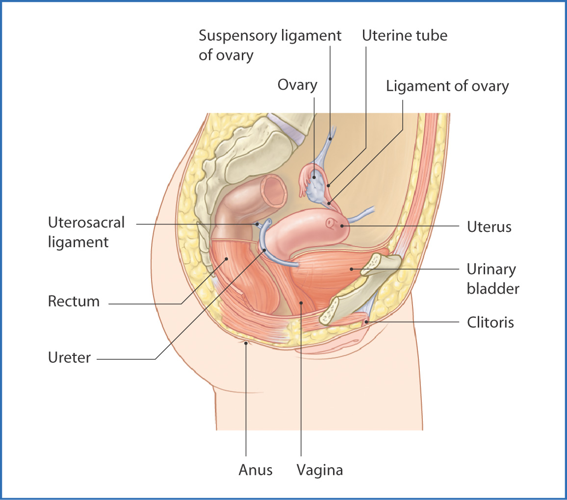
A fold of skin called the clitoral hood covers most of the clitoris, leaving only the tip or nub visible. The rest of the clitoris is a spongy shaft that goes back several inches inside the body. Urethral opening: The urethra is the tube that carries urine from the bladder to the outside of the body. The pelvic floor is a funnel-shaped structure. It attaches to the walls of the lesser pelvis, separating the pelvic cavity from the perineum inferiorly (region which includes the genitalia and anus). In order to allow for urination and defecation, there are a few gaps in the pelvic floor. In comparison to the female pelvis, the male pelvis is narrower. The lesser pelvis in males contains: Distal parts of the urinary and digestive systems: ureter, ...Surface anatomy: Penis (body and glans) and ...Deep perineal pouch: Perineal part of the uret...Superficial perineal pouch: Root of the penis ... Jan 10, 2022 · This diagram depicts Anatomy Female 1024×1111 with parts and labels. Posted in Diagrams, Women Tagged female anatomy, female body, female body diagram, female diagram, female health, female organs, woman anatomy, women. Anatomy. Anatomy: is the study of structures or body parts and their relationships to on another.
Amazon.com: Posterazzi PSTSTK700823H Pelvic floor of human ...
Male pelvis with ligaments, vessels, nerves, pelvic floor and organs, 7-parts - Includes 3B Smart Anatomy This 7 part model of the male pelvis shows in accurate detail how the bones, ligaments, vessels and nerves as well as the pelvic floor muscles and the external genital organs are connected to each other.

Male Pelvis
Sep 07, 2021 · Dog hind leg anatomy. Each hind leg or pelvic limb of a dog consists of half of the pelvic girdle, femur, tibia, fibula, and hind paw. The pelvic girdle of a dog consists of ilium, ischium, pubis, and acetabular bone. Again, the hind paw of a dog consists of tarsal, metatarsals, digits containing phalanges, and the sesamoid bones.
Pelvis Problems | Johns Hopkins Medicine
Human male anatomy scheme. Main pelvis bones - sacrum, ilium, coccyx, pubis, ischium and femur. Vector illustration isolated on a white background. Woman doing exercise with Low Lunge Pose. Woman doing exercise with Low Lunge Pose. Illustration about hip Flexor Stretching. Human anatomy scientific illustrations: female reproductive organ

65 A&P.6.Continuity ideas | reproductive system, human ...
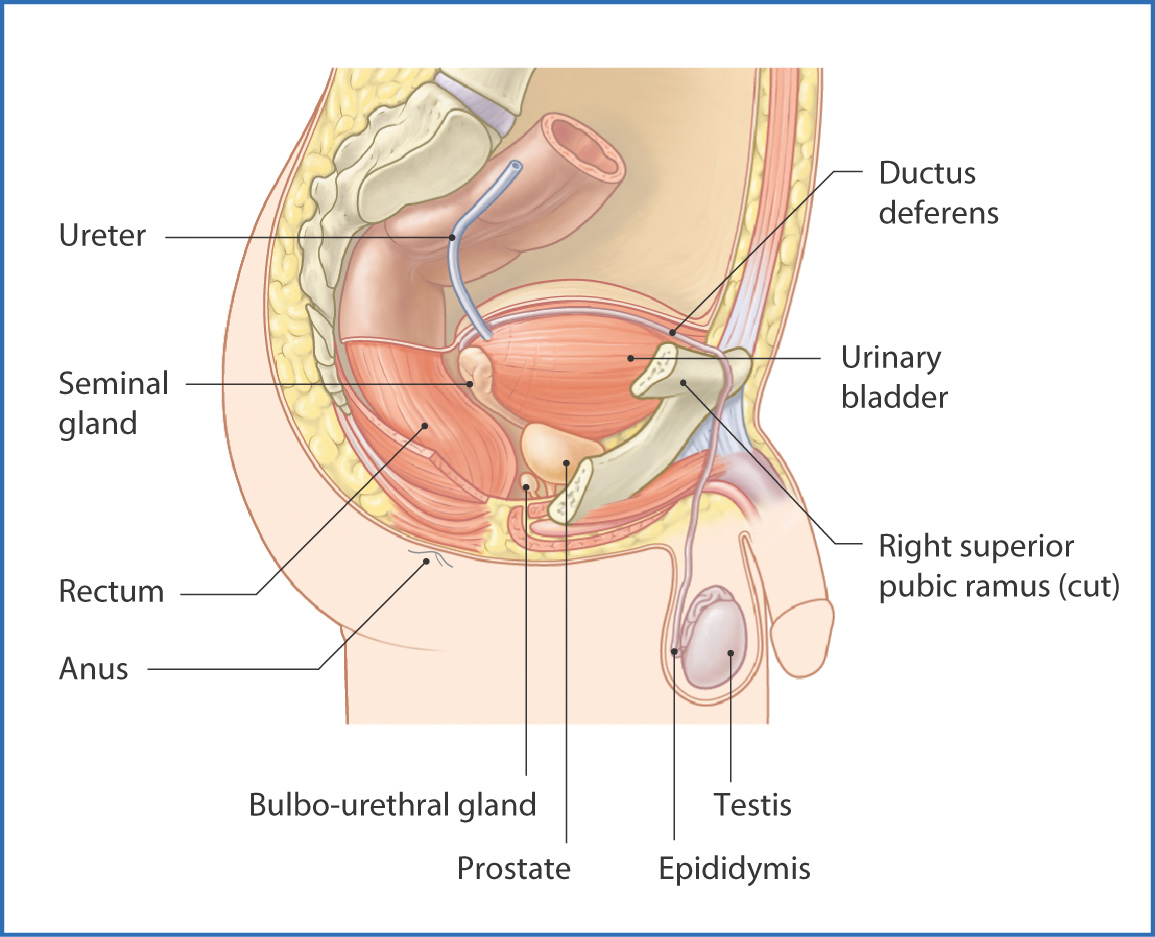
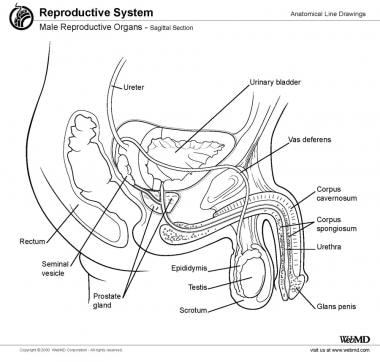
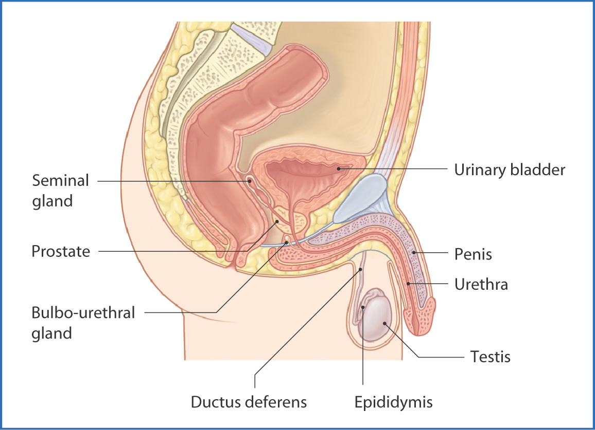
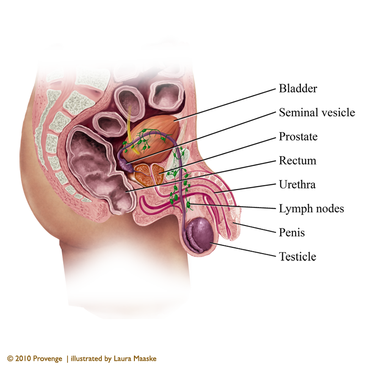
The prostate gland is part of the male reproductive system. It is the size of a walnut and anatomically sits in front of the rectum and between the bladder and the penis. The prostate surrounds the urethra. Sperm are generated in the testes. Tubes called vasa deferens are conduits for sperm that move from the testes to the seminal vesicles.
Pelvic cavity: Anatomical spaces | Kenhub
The muscles of the penis include the corpora cavernosa, two cylinder-like chambers that run down the sides of the penis. Upon arousal, the corpora cavernosa fill with blood, and the penis becomes...
Pelvis and Perineum: Anatomy, vessels, nerves | Kenhub
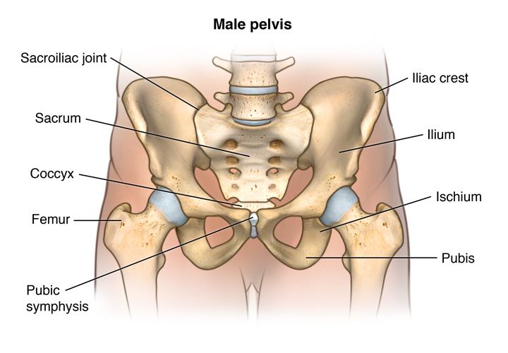
The pelvis is located between the fifth lumbar vertebra and the femoral heads. It forms an irregular bony girdle connecting the lower limbs to the trunk. Anatomy The bony pelvis The bony pelvis is made up of two pelvic bones - the sacrum and the coccyx.

Pelvic Area Diagram Anatomy Of Male Pelvic Area - Human ...
The pelvic cavity, like most spaces in the body, has an inlet and an outlet. For the most part, the pelvic outlet is closed off by the muscles of the pelvic floor (levator ani & coccygeus muscles). The region of the body superficial (caudal in a standing individual) to these muscles and medial to the thighs is known as the perineal region.

Pelvis Anatomical Skeleton Structure. Labeled Vector ...
male pelvis bones Bones The pelvis forms the base of the spine as well as the socket of the hip joint. The pelvic bones include the hip bones, sacrum, and coccyx. The hip bones are composed of...
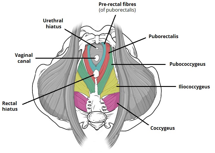
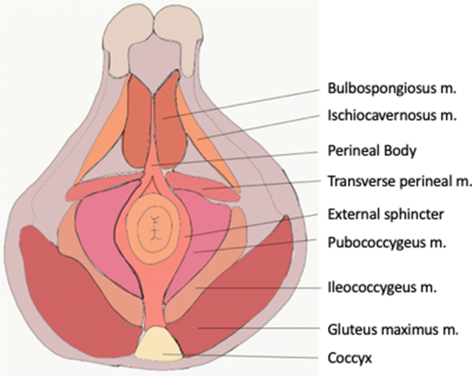
The Pelvic Floor - Structure - Function - Muscles ...
The floor of the pelvis is made up of the muscles of the pelvis, which support its contents and maintain urinary and faecal continence. There are many organs that sit in the pelvis, including much of the urinary system, and lots of the male or female reproductive systems. The skin, tissues and organs in the pelvis are supplied by the ...
5 Facts about the Anatomy of the Pelvic Cavity
May 29, 2018 · Anatomy and function of testes The main function of the testes is producing and storing sperm. They’re also crucial for creating testosterone and other male hormones called androgens.
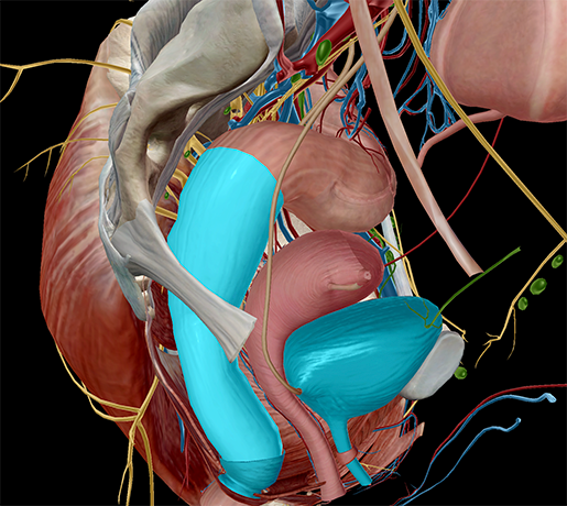
Pelvic Viscera | Basicmedical Key
5 photos of the male chicken anatomy male chicken anatomy. This is also known as the medial compartment of the thigh that consists of the adductor muscles of the hip or.

Male Pelvis, Illustration - Stock Image - C036/6181 - Science ...
The leg of a dog's pelvic limb consists of the tibia and fibula bones. In contrast, the hind paw includes the tarsal bones, metatarsals, and digits that include the three phalanges in each and sesamoid bones. But, how the dog pelvis anatomy is formed? The dog bony pelvis is formed by the ossa coxarum (both sides) and the sacrum bone.

pelvic girdle: In human anatomy, basin-shaped complex of ...
The abdominal viscera occupy the major pelvis; the minor pelvis is the narrower continuation of the major pelvis inferiorly. The inferior pelvic outlet is closed by the pelvic floor. The female pelvis (Figure 1A) has a wider diameter and a more circular shape than that of the male. The wider inlet facilitates head engagement and parturition.
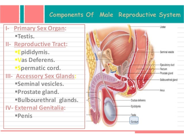
Male Reproductive Organ Anatomy: Overview, Gross Anatomy ...
Introduction The male pelvic floor is a complex structure made up of muscles, ligaments, nerves and fascia. The pelvic floor muscles form part of the pelvic floor and play a critical role in sexual function as well as the maintenance of urinary and faecal continence, + This is a course page funded by Physioplus online learning

Male Pelvis

Pelvis - Wikipedia

MRI of the Male Pelvic Floor | RadioGraphics

The pelvis | Human Anatomy and Physiology Lab (BSB 141)
Atlas of Human Anatomy - 3E

Rear View Male Pelvis Sacrum High Resolution Stock ...

Male Pelvic Floor Muscles and Reproductive Organs ...

Arteries and Veins of Male Pelvis Diagram | Quizlet

5 Facts about the Anatomy of the Pelvic Cavity
Human Papilloma Virus (HPV): Causes, Testing, Treatment ...
2,930 Pelvis Illustrations & Clip Art - iStock
Pelvic Floor Muscle (Kegel) Exercises for Men | Memorial ...
Pelvis and Perineum: Anatomy, vessels, nerves | Kenhub

Male Pelvis
Pelvic anatomy (sagittal view). Each arrow indicates the ...

Module 5: Pelvis Imaging
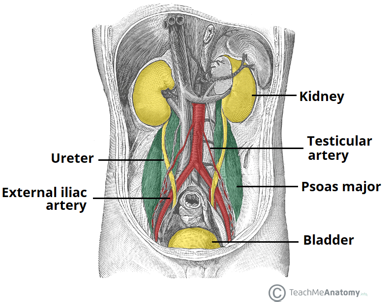
The Ureters - Anatomical Course - Neurovascular Supply ...
Male pelvis viscera
Pelvic Viscera | Basicmedical Key

Male Pelvis with Cancerous Testicle Anatomical Model
Pelvic Viscera | Basicmedical Key
Sagittal section male pelvis | AnatomyTOOL
Male Pelvic Anatomy: Reproductive Organs
Facts About the Spine, Shoulder, and Pelvis
Imaging of chronic male pelvic pain: what the abdominal ...

Male Pelvis Anatomy Diagram | Quizlet

Neurologic Anatomy of the Male Pelvis

3D Anatomical model of the male pelvis (3B Scientific) from ...
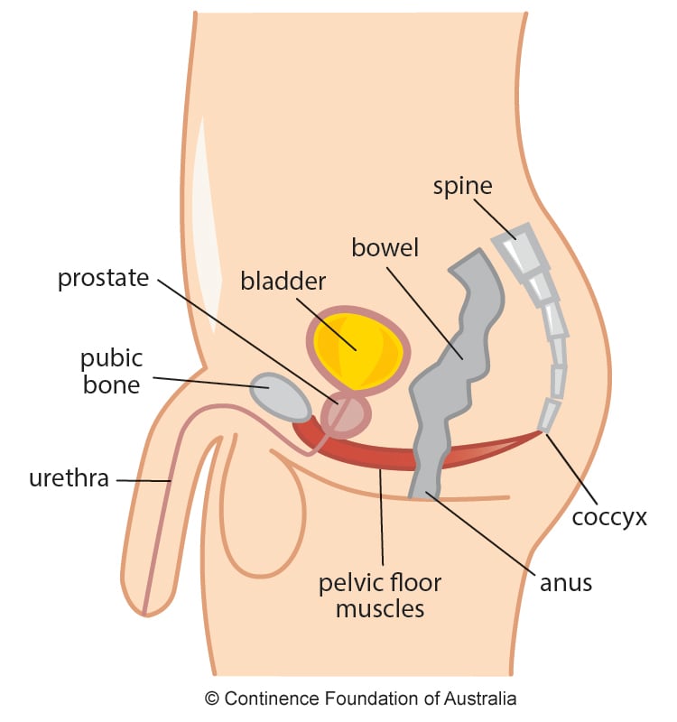
Male Pelvic Floor | Exercises | Continence Foundation of ...


Comments
Post a Comment