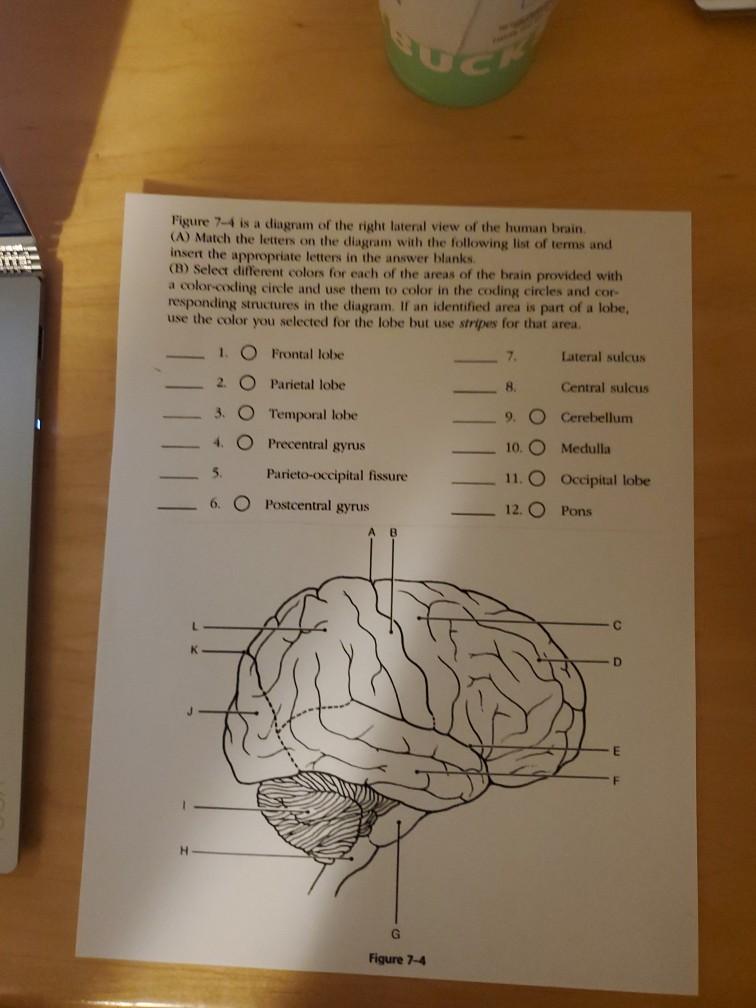
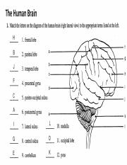
43 match the letters on the diagram of the human brain
Match Gravity HUMAN BRAIN- RIGHT LATERAL VIEW Click card to see definition 👆 A = POSTCENTRAL GYRUS B = PARIETAL LOBE C = PARIETO-OCCIPITAL SULCUS D = OCCIPITAL LOBE E = CEREBELLUM F = PRECENTRAL GYRUS G = CENTRAL SULCUS H = FRONTAL LOBE I = LATERAL SULCUS J = TEMPORAL LOBE K = PONS L = MEDULLA Click again to see term 👆 1/102 Previous ← Next → Figure 7-4 is a diagram of the sagittal view of the human brain. First, match the letters on the diagram with the list of terms and insert the appropriate letter in each answer blank. Then, color the brain-stem areas blue and the areas where cerebrospinal fluid is found yellow. 1. Cerebrum 8. Pituitary gland (Endocrine gland called Master Gland)
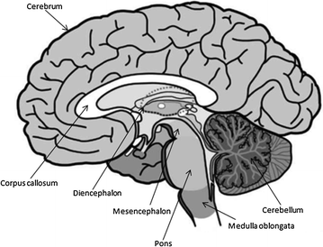
Figure 12.2 Embryonic development of the human brain. (e) Adult neural canal regions (d) Adult brain structures (a) Neural tube (c) Secondary brain vesicles (b) Primary brain vesicles Anterior Diencephalon (rostral) Posterior (caudal) Spinal cord Central canal Cerebellum Brain stem: medulla oblongata Brain stem: pons Brain stem: midbrain
Match the letters on the diagram of the human brain
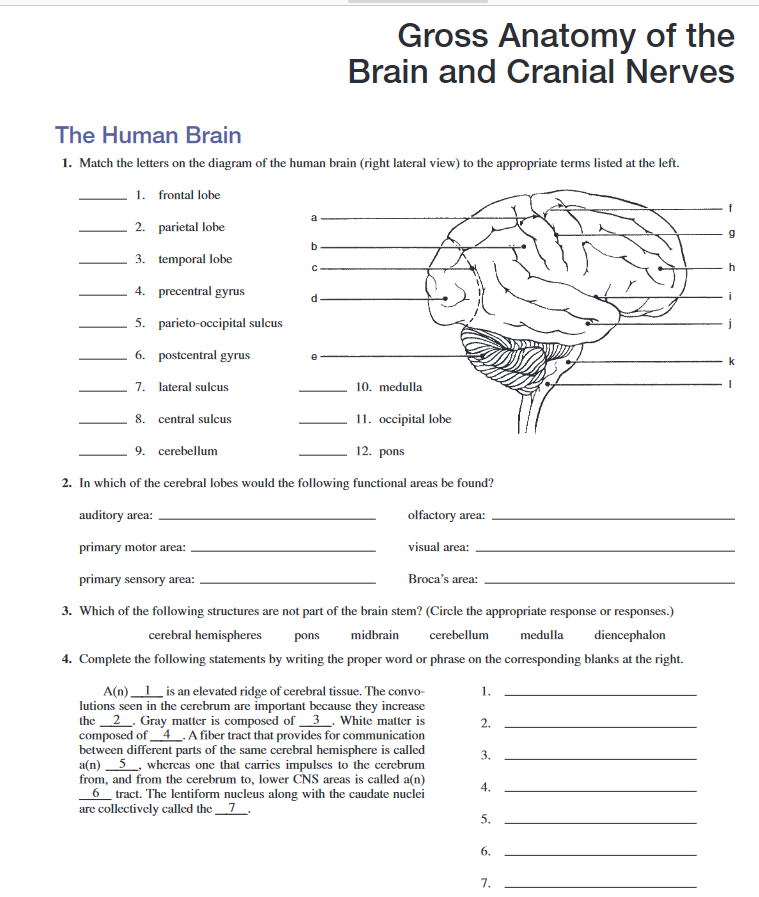
Match the letters on the diagram of the human brain (right lateral view) to the appropriate terms listed at the left. H 1 . frontal lobe B 2. parietal lobe J 3. temporal lobe F 4 . precentral gyrus C 5. parieto-occipital sulcus A 6. postcentral gyrus I 7. lateral sulcus G 8. central sulcus E 9. cerebellum L 10. medulla D 11. occipital lobe K 12. pons 2. the diagram at the right. Multiple sclerosis (MS) is a potentially disabling disease of the brain and spinal cord (central nervous system). In MS, the immune system attacks the protective sheath (myelin) that covers nerve fibers and causes communication problems between your brain and the rest of your body. Match the letters on the diagram of the human brain (Gright lateral view) to the appropriate terms listed at the left: lateral view) to 1. frontal lobe 2. parietal lobe 3. temporal lobe 4. precentral gyrusd 5. parieto-occipital sulcus 6. postcentral gyrus 7. lateral sulcus 8. central sulcus 9. cerebellum 10, medulla 11, occipital lobe 12. pons
Match the letters on the diagram of the human brain. Of all the human body systems, the nervous system is the most complicated system in the body. The brain is the central part of the nervous system. It is an intriguing organ, that has been studied right from the time it develops in the fetus. The human brain weighs about 1.5 kg in adults. The Human Brain I. Match the letters on the diagram of the human brain (right lateral view) to the appropriate terms listed on the left. H 1. frontal lobe B 2. parietal lobe J 3. temporal lobe F 4. precentral gyrus C 5. parieto-occipital sulcus A 6. postcentral gyrus I 7. lateral sulcus G 8. central sulcus E 9. cerebellum L 10. The gross anatomy of the human brain can vary, but since the development of the brain is strictly coded by genes, many of the notable structures have an uniformed appearance and are the same in the brain of every human being. These structures are in the form of the gyri and sulci, and the way they are positioned to each other defines the names ... First, match the letters on the diagram with the following list of terms and insert the appropriate letters in the answer blanks. Then, select different colors for each of the areas of the brain provided with a color-coding circle and use them to color in the coding circles and corresponding structures in the diagram.
2. Match the letters on the diagram of the human brain (right lateral view) to the appropriate terms listed at the left: 1. 2. 3. 4. 5. 6. 7. 8. 9. frontal lobe parietal lobe temporal lobe precentral gyrus parieto-occipital sulcus postcentral gyrus lateral sulcus central sulcus cerebellum 10. medulla oblongata 11. occipital lobe 12. pons 3. BI 335 - Advanced Human Anatomy and Physiology Western Oregon University Figure 4: Mid-sagittal section of brain showing diencephalon (includes corpus callosum, fornix, and anterior commissure) Marieb & Hoehn (Human Anatomy and Physiology, 9th ed.) - Figure 12.10 Exercise 2: Utilize the model of the human brain to locate the following structures / landmarks for the Plasticity The brain's ability to recover from brain/nerve damage by possibly creating new pathways for previous messages Action potential This allows messages to flow from neuron to neuron as an electrical charge is created when positively charged sodium ions flow into a neuron and flows out as positively charged potassium charges. First, match the letters on the diagram with the following list of terms and insert the appropriate letters in the answer blanks. Then, select different colors for each of the areas of the brain provided with a color-coding circle and use them to color in the coding circles and corresponding structures in the diagram.
The processing power and memory capacity necessary to match general intellectual performance of the human brain are estimated. Based on extrapolation of past trends and on examination of technologies under development, it is predicted that the required hardware will be available in cheap machines in the 2020s. retina converts light into electrical impulses that are sent to the brain through the optic nerve. Vitreous gel: The vitreous gel is a transparent, colorless mass that fills the rear ... parts of the eye, eye diagram, vitreous gel, iris, cornea, pupil, lens, optic nerve, macula, retina ... Match the letters on the diagram of the human brain (right lateral view) to the appropriate terms listed at the left. h 1. frontal lobe b 2. parietal lobe J 3. temporal lobe f 4. precentral gyrus c 5. parieto-occipital sulcus a 6. postcentral gyrus i 7. lateral sulcus g 8. central sulcus e 9. cerebellum l 10. medulla d 11. occipital lobe k 12. pons 2. Labeled brain diagram. First up, have a look at the labeled brain structures on the image below. Try to memorize the name and location of each structure, then proceed to test yourself with the blank brain diagram provided below. Labeled diagram showing the main parts of the brain.
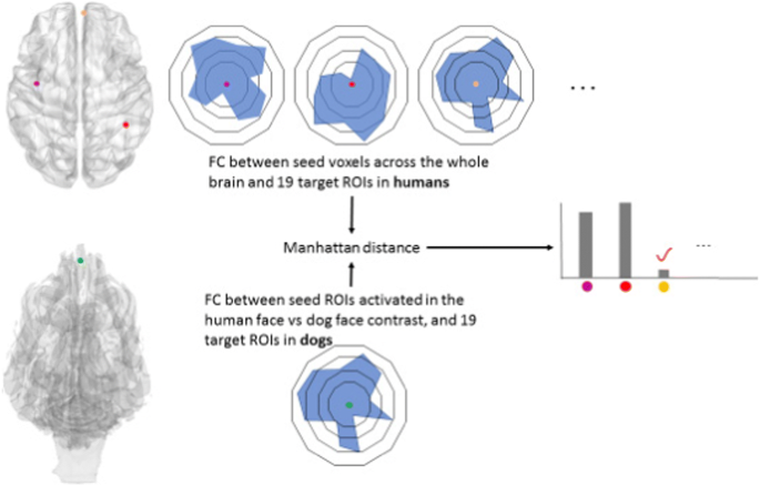
Separate brain areas for processing human and dog faces as ...
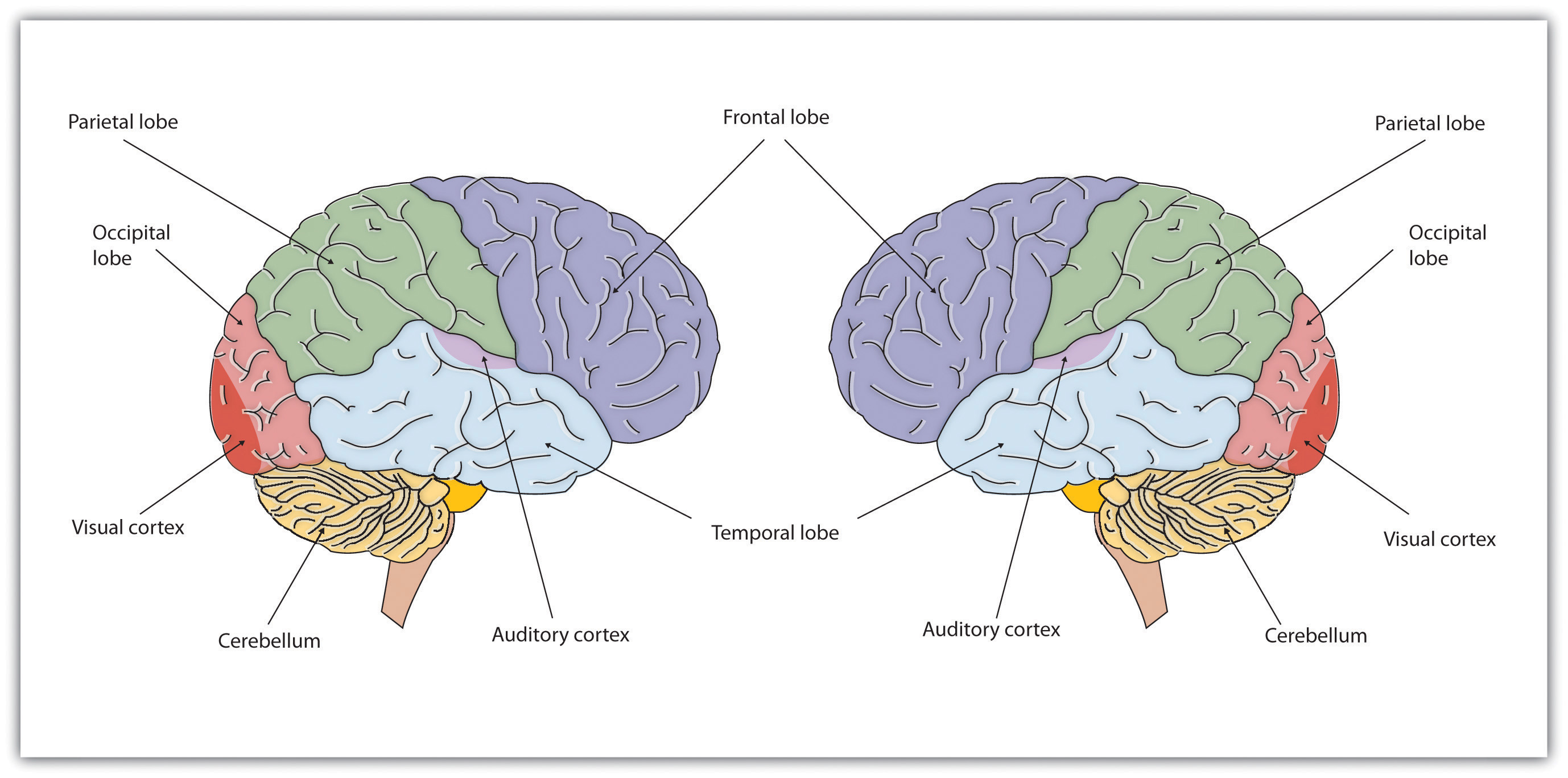
You will be asked to identify the major parts of the brain on a diagram. Items are limited to the following: cerebrum. cerebellum. pons. medulla oblongata. brain stem. frontal lobe. parietal lobe.
Basics of Brain Computer Interface | SpringerLink
The midbrain (or mesencephalon ), located near the very center of the brain between the interbrain and the hindbrain, is composed of a portion of the brainstem. The hindbrain (or rhombencephalon) consists of the remaining brainstem as well as our cerebellum and pons.

Learning Resources Cross-section Brain Model - 2 Pieces, Ages 7+ Brain Anatomy Model, Brain Functions Model, Human Anatomy for Kids, Foam Brain Model
The figure below is sagittal view of the human brain. First match the letters on the diagram with the following list of terms and insert the appropriate letter in each answer blank. Then color the brain-stem areas blue and the areas where cerebrospinal fluid is found yellow. 1. Cerebellum 4. Pituitary gland 7. Corpus callosum 10. Medulla ...
Solved XX Figure 7-4 is a diagram of the right lateral view ...
amazing revision guide name per. nervous system study guide introduction: the nervous system is the master coordinating system of the body. every thought,
Assignment 15 pg 165.pdf - Review Sheet 10 165 | Course Hero
The diagram of the brain is useful for both Class 10 and 12. It is one among the few topics having the highest weightage of marks and is frequently asked in the examinations. A well-labelled diagram of a human brain is given below for further reference.

The diagram below shows the human brain.Unscramble the words ...
Nervous system. Base your answer to the following question on the diagram below illustrating one type of cellular communication and your knowledge of biology. In region F, there is a space between nerve cells C and D. Cell D is usually stimulated to respond by.

In the diagram of the lateral view of the human brain, parts are indicated by alphabets. Select the answer in which these alphabets have been correctly matched with the parts which they indicate.
The brain is the ultimate organ of adaptation. It takes in information and orchestrates complex behavioral repertoires that allow human beings to act in sometimes marvelous, sometimes terrible ways. Most of what people think of as the "self"—what we think, what we remember, what we can do, how we feel—is acquired by the brain from the experiences that occur after birth.

A Pattern Recognition Theory of Mind - Forte Labs
1. Match the letters on the diagram of the human brain (right lateral view) to the appropriate terms listed at the left. 1. frontal lobe 2. parietal lobe 3. temporal lobe 4. precentral gyrus 5. parieto-occipital sulcus 6. postcentral gyrus 7. lateral sulcus 10. medulla 8. central sulcus 11. occipitallobe 9. cerebellum 12. pons 2.
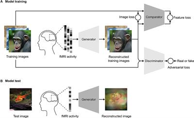
Frontiers | End-to-End Deep Image Reconstruction From Human ...
Created Date: 4/30/2013 4:05:46 PM

A&P Lab Exercises 17&19 Flashcards | Quizlet
The height of the human brain is about 3.6 inches and it weighs about 4 to 5 lbs at birth and 3 lbs in adults. The total surface area of the cerebral cortex is about 2,500 cm2 and when stretched, it will cover the area of a night table. The brain is composed of 77 to 78% water and 10 to 12% lipids. It contains 8% proteins 1% carbohydrates, 2% ...

a&p_lab_ex_19 - NAME LAB TIME/DATE _ Gross Anatomy of the ...
Match the letters on the diagram of the human brain (Gright lateral view) to the appropriate terms listed at the left: lateral view) to 1. frontal lobe 2. parietal lobe 3. temporal lobe 4. precentral gyrusd 5. parieto-occipital sulcus 6. postcentral gyrus 7. lateral sulcus 8. central sulcus 9. cerebellum 10, medulla 11, occipital lobe 12. pons

The Situated Brain (Section 2) - Culture, Mind, and Brain
the diagram at the right. Multiple sclerosis (MS) is a potentially disabling disease of the brain and spinal cord (central nervous system). In MS, the immune system attacks the protective sheath (myelin) that covers nerve fibers and causes communication problems between your brain and the rest of your body.
Brain Activity Language High Resolution Stock Photography and ...
Match the letters on the diagram of the human brain (right lateral view) to the appropriate terms listed at the left. H 1 . frontal lobe B 2. parietal lobe J 3. temporal lobe F 4 . precentral gyrus C 5. parieto-occipital sulcus A 6. postcentral gyrus I 7. lateral sulcus G 8. central sulcus E 9. cerebellum L 10. medulla D 11. occipital lobe K 12. pons 2.

The diagram given below is an external view of the human ...
a&p_lab_ex_19 - NAME LAB TIME/DATE _ Gross Anatomy of the ...
The diagram shows a section of the human brain. Answer the ...

Physical principles of brain–computer interfaces and their ...

Brain Basis of Human Social Interaction: From Concepts to ...

The Brain Basis of Language Processing: From Structure to ...
Chapter 3 – Neuron and the Brain – Psychology
Lab 12 - Question 1 of 8 Match the letters on the diagram of ...

Brain Basis of Human Social Interaction: From Concepts to ...
Internal carotid artery: Anatomy, segments and branches | Kenhub
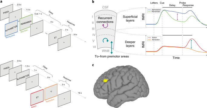
Layer-dependent activity in human prefrontal cortex during ...
![PDF] Audiovisual Integration of Letters in the Human Brain ...](https://d3i71xaburhd42.cloudfront.net/95de555bcab9d6424aba64ec4697e30e1b6e02ac/4-Figure4-1.png)
PDF] Audiovisual Integration of Letters in the Human Brain ...

Precision biomarkers for mood disorders based on brain ...

Ageâ€related differences in autism: The case of white matter ...

Lab 12 - Question 1 of 8 Match the letters on the diagram of ...

A comes before B, like 1 comes before 2. Is the parietal ...

Dynamic Stimulation of Visual Cortex Produces Form Vision in ...
Are Computers Already Smarter Than Humans? | Time

Ch 14-16, 19 Flashcards | Quizlet
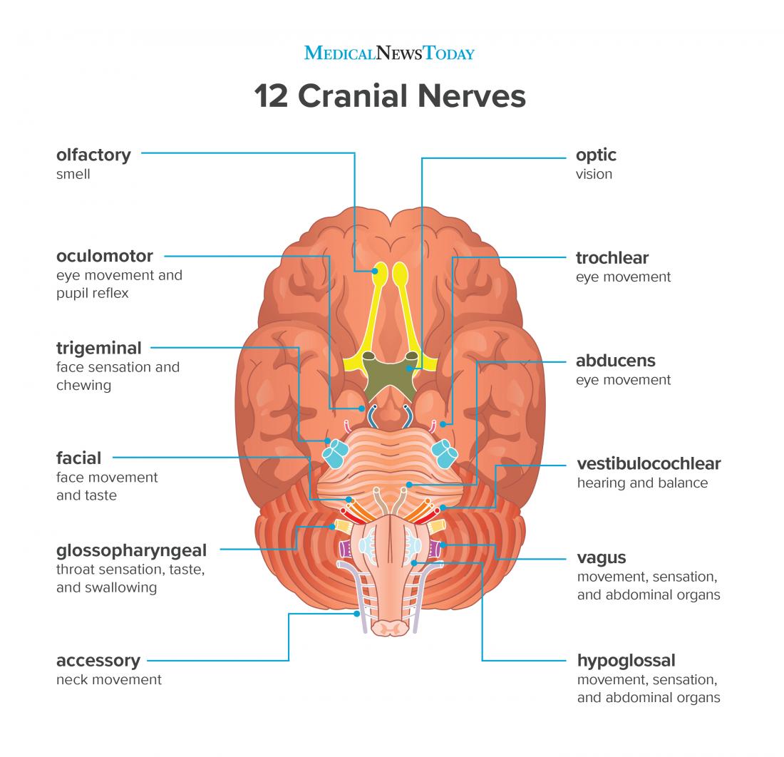
What are the 12 cranial nerves? Functions and diagram

Brain and Cognitive Development - Stiles - - Major Reference ...

A brief history of the brain | New Scientist
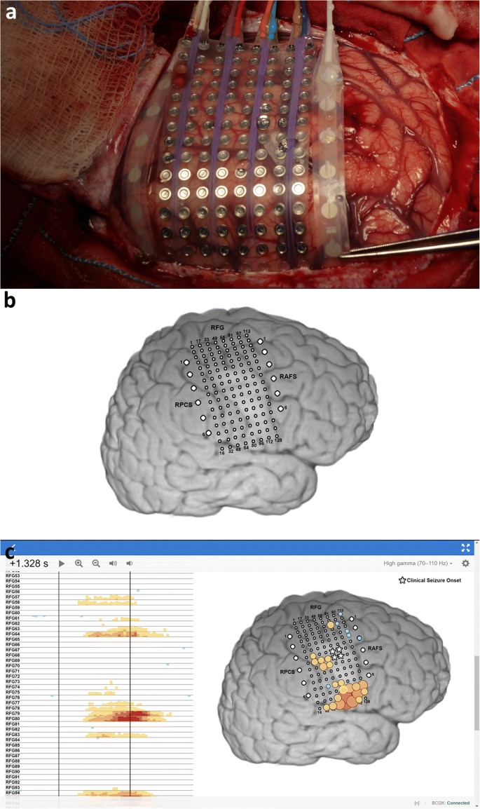
How to map the brain
The Potential for a Speech Brain–Computer Interface Using ...

a&p_lab_ex_19 - NAME LAB TIME/DATE _ Gross Anatomy of the ...

The Brain Basis of Language Processing: From Structure to ...
a&p_lab_ex_19 - NAME LAB TIME/DATE _ Gross Anatomy of the ...

In the given diagram of section of brain, different parts are ...
Solved Gross Anatomy of the Brain and Cranial Nerves The ...

Label The Parts Of The Brain Worksheet Worksheets For All ...


Comments
Post a Comment