43 sheep heart diagram labeled
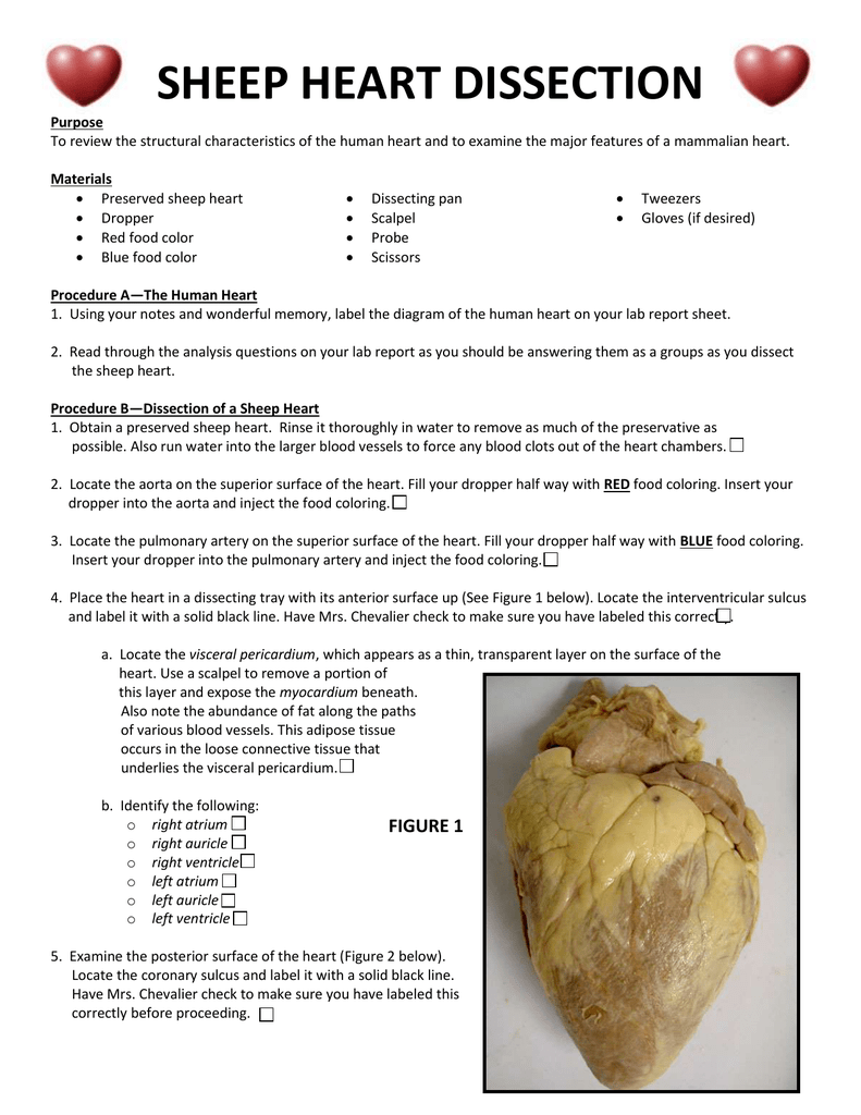
By studying the sheep’s anatomy, you can learn how your own heart pumps blood through your body, thereby keeping you alive! Use this sheep heart dissection guide in a lab for high school students. You can also look at the labeled pictures to get an idea of what the heart looks like (that’s especially helpful for younger students). Procedure B—Dissection of a Sheep Heart. 1. Obtain a preserved sheet heart. Rinse it in water thoroughly to remove as much of the preservative as possible. 10. Locate the parts of the heart (labeled on the human heart diagram of your lab report) on a dissectible heart model.
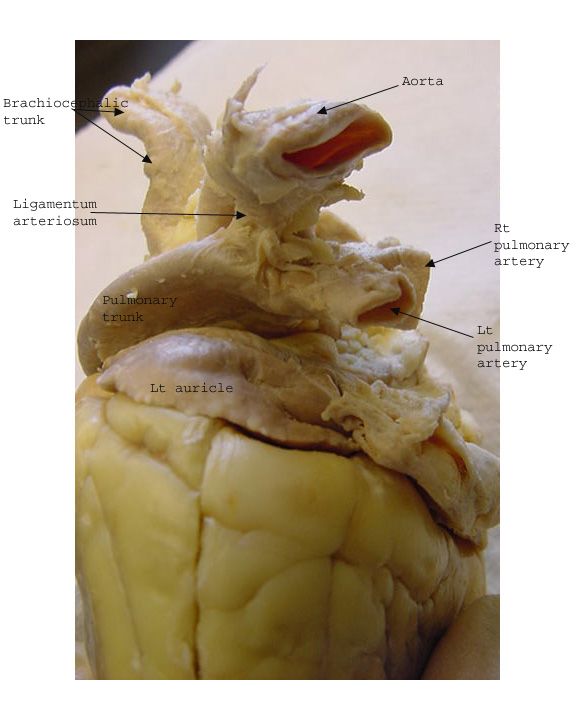
Sheep Heart Dissection Procedure (Day 2) – you will be cutting the heart open today! a. Review the outer part of the heart and make sure you know where these structures are: left and right ventricle, left and right atrium, pulmonary artery, pulmonary veins, aorta, coronary artery, apex, superior and inferior vena cava. b. Locate the pulmonary ...
Sheep heart diagram labeled
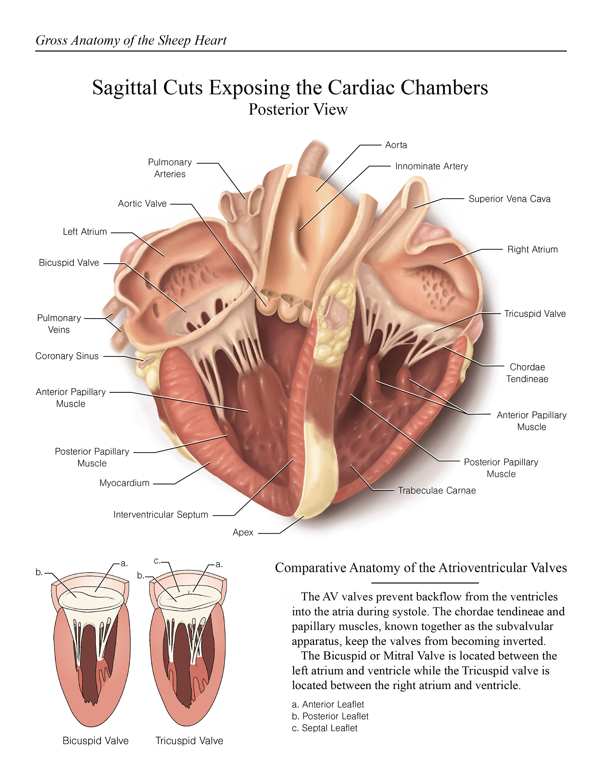
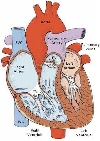
Image: Labeled diagram of the heart showing the aortic valve anatomically located between the left ventricle and aorta. Blood Flow Through the Heart. Now that we have a good understanding of the 4 chambers and valves of the heart, there are only 4 more main structures we will discuss. Label The Parts Of The Heart. depts.washington.edu | Having the heart diagram for studies … Labeled Heart Diagram Download. hatrc.org/library | It is an easy to download template … Label parts of the heart Drag and drop the labels to the correct parts indicated on the heart... (a) Anterior view of the external heart C' 2019 Pearson Education. Aort'c arch Ligamentum arteriosum Left pulmonary artery Left pulmonary ve ns Auricle of left atrium Circumflex artery Left coronary artery (in atrioventricular sulcus) Great cardiac vein Left ventricle Anterior interventricular artery (in anterior interventricular sulcus) Apex
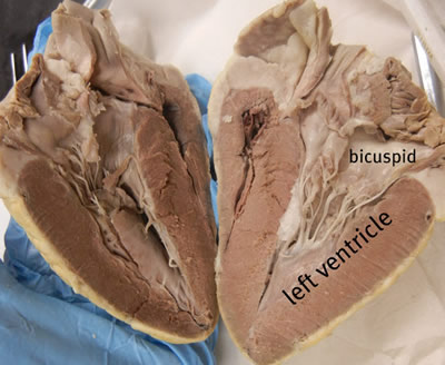
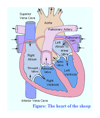
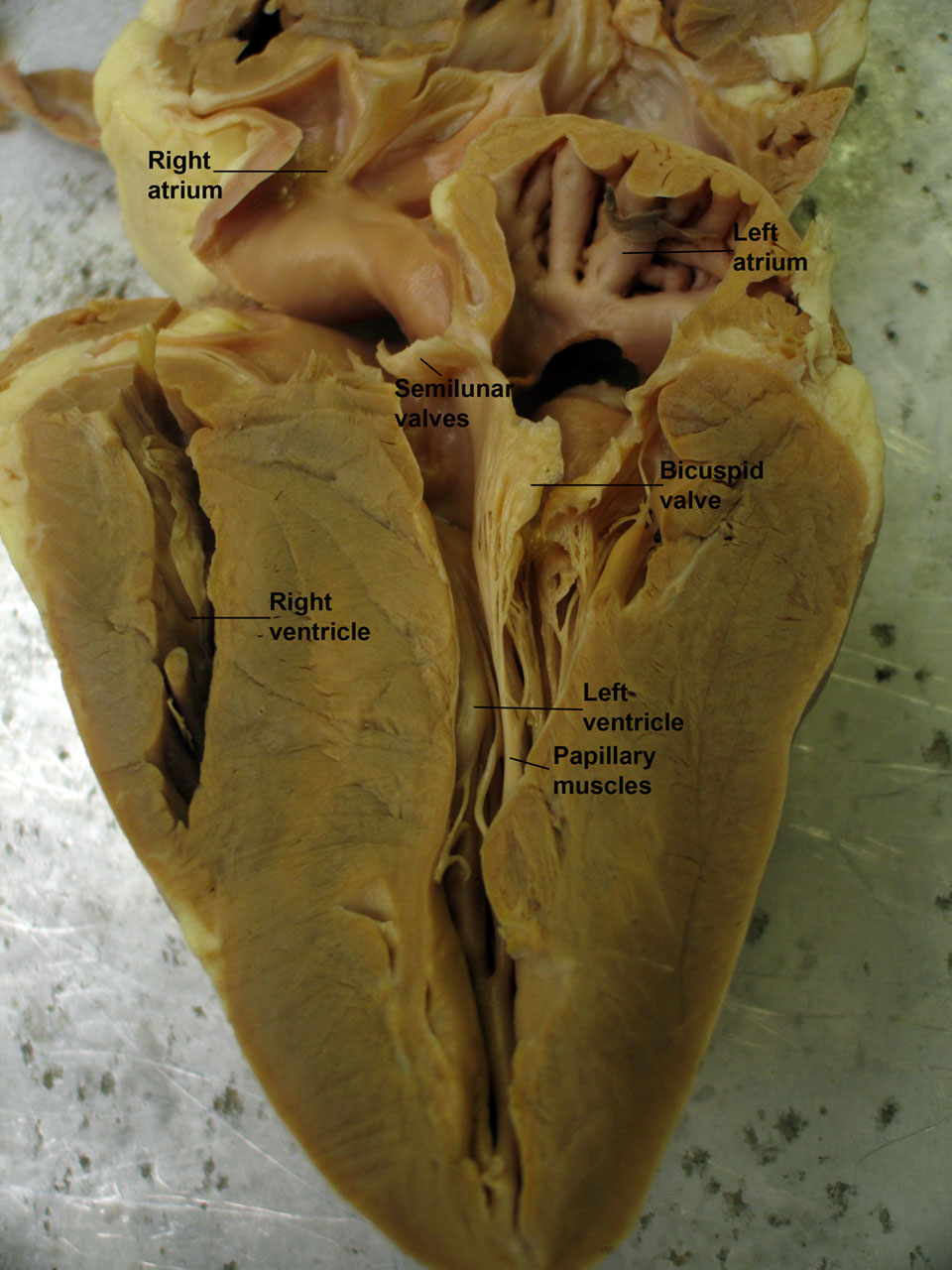
Sheep heart diagram labeled. Sheep heart anatomy labeled. Article by minted peach. Sheep heart anatomy including atria ventricles vena cava aorta pulmonary artery vein tricuspid valve bicuspid valve aortic and pulmonic valves trab. Bio 117 Lab 1 Sheep Heart Dissection Exterior Diagram Quizlet. Take this practice quiz that covers information related to the sheep heart & the heart model. It is intended for use as a supplemental study aid. As is the case in the lab practical, each correct answer counts. So, make sure you learn from the feedback. Questions and Answers. 1. Whole Sheep Heart Diagram Quizlet. 2114 Anatomy And Physiology Ii Open Virtual Laboratory. Anatomy Of The Sheep Heart. Sheep Heart Anterior View Diagram Quizlet. Sheep Heart (right side). This image shows a close-up view of the inside of the right ventricle. Blood from the right atrium enters the right ventricle through the one-way tricuspid valve.
Sheep Heart Dissection Lab -Loudoun County Public Schools Sheep Heart Dissection Procedure (Day 2) - you will be cutting the heart open. your textbook and/or your notes and heart diagram worksheet, label the diagram of the human heart on your lab report sheet. 1. Using your notes and wonderful memory, label the diagram of the human heart on your lab report sheet. 2. Read through the analysis questions on your lab report as you should be answering them as a groups as you dissect . the sheep heart. Procedure B—Dissection of a Sheep Heart. 1. Obtain a preserved sheep heart. Sheep. Heart Dissection Lab. Introduction Mammals have four-‐chambered hearts and double circulation. The heart of a bird or mammal has Procedure—External Anatomy Most heart diagrams show the left atrium and ventricle on the right side of the diagram. Imagine the heart in the body of a... Sheep Heart Dissection External Structures And Blood Vessels Youtube. Sheep Heart Dissection Guide Pdf Diagram Notasdecafe Co. Superior And Inferior Vena Cava Sheep Heart. Midterm Lab Exam A Pii Sheep Heart Flashcards Quizlet.
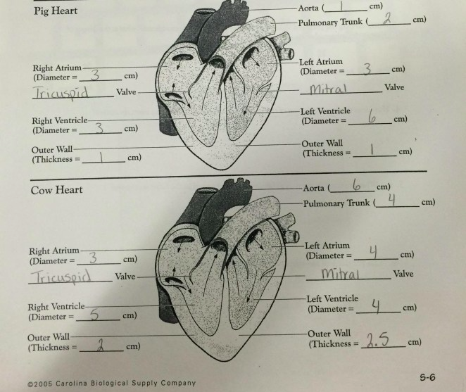
Sheep Heart Diagram Labeled. Last modified: 2021-09-19. ANTERIOR SHEEP HEART - Biology Forums Gallery. Anat lab 2 - Anatomy & Physiology 223 with Cummings at ... Lab 3: heart and vessels at Southern Illinois University ... Parts we have to know.Sorry it's not so clear!I tried to focus it. but i mean you can still see it.just a little blurry that's all..Study Study... The sheep hearts contain Carosafe, which is a preservative.You may want to use gloves when handling the sheep heart, although it is not necessary. Matching game: 1. Make sure there are three bags each of Crisco and sugar, labeled with the number of grams. in each bag. Sheep Heart Diagram Labeled. Ditulis JupiterZ Senin, 04 Januari 2021 Tulis Komentar Edit. 28 Cow Heart Diagram Anatomy And Physiology The Heart On The Cutting Edge Sheep Heart Dissection Carolina Com
Day 10 - Heart Dissection powerpoint
First, it has a few nicely labeled images you can use to test your knowledge. It has a basic diagram/sequence of blood flow through the heart. Finally, at the bottom of the page there are three practice documents: External anatomy: label the valves (.pdf), Internal anatomy: label the right side (.pdf) Internal anatomy: label the left side (.pdf)

Sheep Heart Images
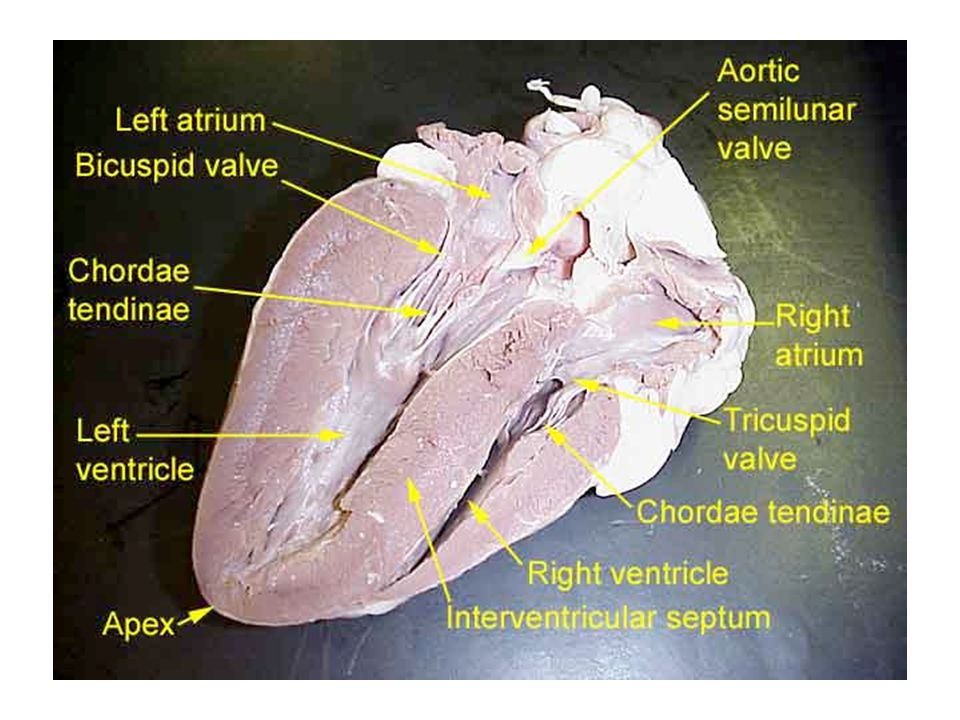
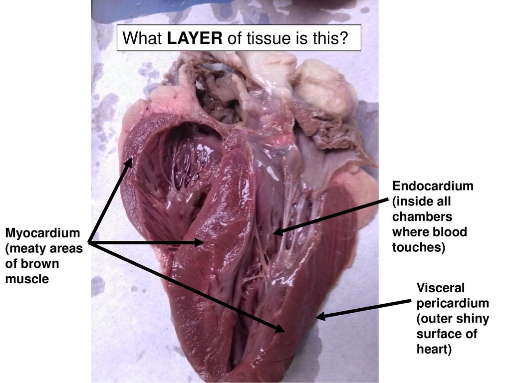
This page contains photos of the sheep heart dissection. All the major vessels are represented, many are labeled with colored pencils so that you can see exactly where each The heart can be confusing because it is not perfectly symmetrical. Students often confuse the left and the right side of the heart.

Semilunar valve Definition and Examples - Biology Online ...
Lab guide outlining the procedure for dissecting the sheep's heart. It includes photos to diagram where major vessels are and where incisions should be made to view internal Checkpoint: Make sure you know the location of each of the following before continuing to the internal anatomy of the heart

2: Diagram of the sheep lung divided into segments. These ...
2 atria, 2 ventricles, completely closed circulatory system with no mixing of blood. Base. the wide upper or anterior end of the heart, where the aorta and vena cava are located. apex. somewhat blunt tip of the heart, composed entirely of the left ventricle. musculi pectinati. parallel ridges in the walls of the atria of the heart.

Sheep Heart Dissection - Lessons - Blendspace
Sheep Heart Diagram. Posted on December 21, 2018December 21, 2018. Sponsored links. Related posts: 6.7 Cummins Belt Diagram. 89 Ford F150 Wiring Diagram. Labelled Diagram Of Bacteria.

Heart Anatomy | Anatomy and Physiology II
Mar 06, 2021 · Start studying Sheep heart: labeled. Learn vocabulary, terms, and more with flashcards, games, and other study tools.
Heart Anatomy/Heart Dissections/Heart Labeling ...
Sheep Heart Unlabeled. Sheep Heart Leader-lined. Sheep Heart Labeled. San Diego Mesa College 7250 Mesa College Drive San Diego, CA 92111-4998 Student Support San Diego Community College District San Diego City College San Diego Mesa College San Diego Miramar College San Diego Continuing Education.
Title: Sheep Heart Dissection Objective: -To Practice ...
This online quiz is called Sheep Heart Labeling comparative anatomy, heart diagram, sheep heart. Sheep Heart Labeling. a quiz by renwickrebecca. • 4,656 plays.

Image result for sheep heart labeled | Heart anatomy, Medical ...
This page contains photos of the sheep heart dissection. All the major vessels are represented, many are labeled with colored pencils so that you can see. In this lab guide, students are given instruction on how to remove the dura mater, and locate the main structures of the external and internal brain.

ANTERIOR SHEEP HEART - Biology Forums Gallery
In this interactive, you can label parts of the human heart. Drag and drop the text labels onto the boxes next to the heart diagram. If you want to redo an answer, click on the box and the answer will go back to the top so you can move it to another box. If you want to check your answers, use the Reset...
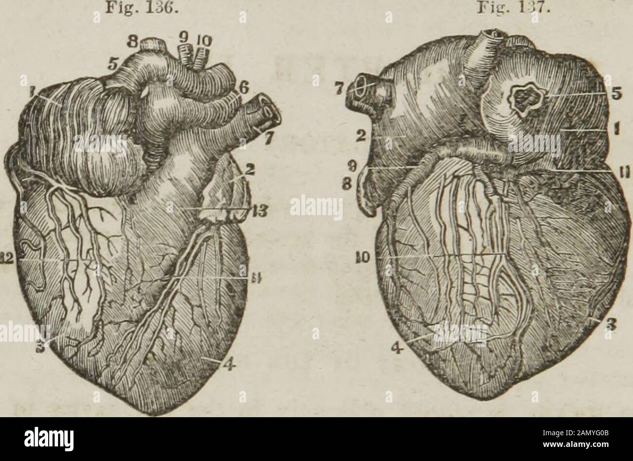
Anatomy and physiology : designed for academies and families ...
Start studying sheep heart labeling. Learn vocabulary, terms and more with flashcards, games and other study tools.
Anatomy & Physiology
Sheep Heart Dissection. What are the chambers, valves, and blood vessels of the heart? Identify the right and left sides of the heart Obtain a sheep heart and place the heart in a dissecting tray. Position your heart in the tray so that it matches the diagram below.
Lab 04 - Heart Anatomy
SHEEP HEART DISSECTION Purpose To review the structural characteristics of the human heart and to examine the major features of a mammalian heart. Period _____ Please label the diagram of the human heart below.
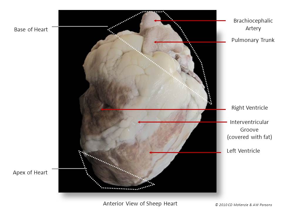
Anterior View of Sheep Heart - ppt video online download
Heart of a sheep diagram online drga and drop. Play here >>>. These diagrams are also like science games and teachers can use them as group games in the classroom to test and see how good students are in labelling science diagrams.
Answered: do you have a diagram of the sheep… | bartleby
Family. Unfollow. Labeled Sheep Heart Picture #4. Done.
Day 10 - Heart Dissection powerpoint
sheep heart. Observation: External Anatomy Most heart diagrams show the left atrium and ventricle on the right side of the diagram. Imagine the heart in the body of a person facing you. The left side of their heart is on their left, but since you are facing them, it is on your right. 1. Identify the right and left sides of the heart. Look ...
Sheep Heart. - ppt download
4. Compare the structures of the sheep heart with those of the human heart. 5. Know the path of blood through and out of the heart Materials Preserved sheep heart Dissecting tray and instruments Vinyl dissecting aprons Disposable gloves Anatomy & Physiology / Revealed, Version 2.0 CD-ROM Human heart model

Sheep Heart Images
Materials • Sheep heart specimen • Dissection tray or Styrofoam meat tray • Dissection kit with Interesting Facts • Most heart diagrams show the left atrium and ventricle on the right side of the diagram. Anatomy www. enchantedlearning. com/subjects/ana tomy/heart labelinterior/label. shtml.
Sheep Heart Dissection Lab
Can someone please help me label this sheep heart diagram? Thank you so much! Or at least.

internal sheep heart labeling 1 Diagram | Quizlet
Sheep Heart from anatomycorner.com. See more ideas about heart anatomy, heart diagram, anatomy and physiology. Precautions to prevent heart diseases. One with labels attached, and one blank diagram with the labels at the bottom for students to complete themselves.

BIO202-Sheep Heart
Anatomy Lab: Sheep Heart Diagram - Quizlet. 2 hours ago 2 atria, 2 ventricles, completely closed circulatory system with no mixing of blood. Base. the wide upper or anterior end of the heart, where the aorta and vena cava are located. apex. somewhat blunt tip of the heart, composed entirely of the left...

Sheep Heart Images
(a) Anterior view of the external heart C' 2019 Pearson Education. Aort'c arch Ligamentum arteriosum Left pulmonary artery Left pulmonary ve ns Auricle of left atrium Circumflex artery Left coronary artery (in atrioventricular sulcus) Great cardiac vein Left ventricle Anterior interventricular artery (in anterior interventricular sulcus) Apex
Dissected Quotes. QuotesGram
Label The Parts Of The Heart. depts.washington.edu | Having the heart diagram for studies … Labeled Heart Diagram Download. hatrc.org/library | It is an easy to download template … Label parts of the heart Drag and drop the labels to the correct parts indicated on the heart...
Copy of Virtual Sheep Heart Dissection
Image: Labeled diagram of the heart showing the aortic valve anatomically located between the left ventricle and aorta. Blood Flow Through the Heart. Now that we have a good understanding of the 4 chambers and valves of the heart, there are only 4 more main structures we will discuss.
Sheep Heart Dissection on Behance

Procedure B Dissection Of A Sheep Heart - Human Anatomy ...

Sheep Heart Dissection

Labeled Sheep Heart Picture #3 | Alexandria | Flickr

Heart (Sheep) External Anatomy - YouTube

Heart Structure | BioNinja
www.robert-pace.com » Blog Archive » Zoology Dissection ...
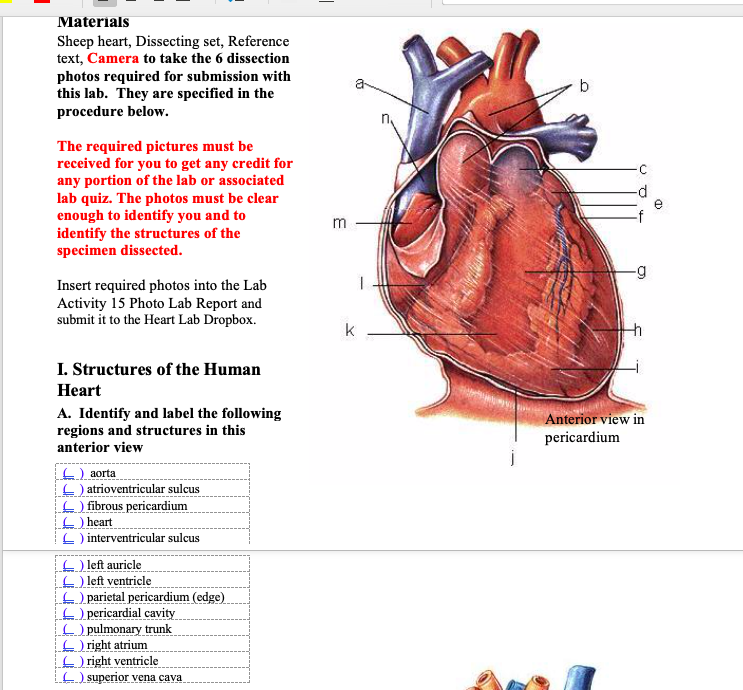
Solved Materials Sheep heart, Dissecting set, Reference ...

Heart Dissection! | Aricka's Anatomy Blog

Quiz: Can You Identify These Parts Of A Sheep's Heart ...
Untitled

Heart Dissection | Carolina.com

Sheep heart dissection: external structures and blood vessels

Heart Structure | BioNinja

Sheep Heart Dissection - Part 2 - YouTube
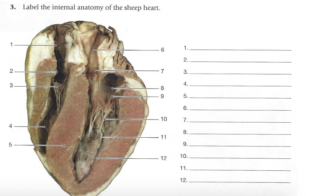
Solved 3. Label the internal anatomy of the sheep heart. 6 ...

Sheep Heart Dissection

Heart Dissection | bulb
Sheep Heart Dissection


Comments
Post a Comment