40 diagram of villi
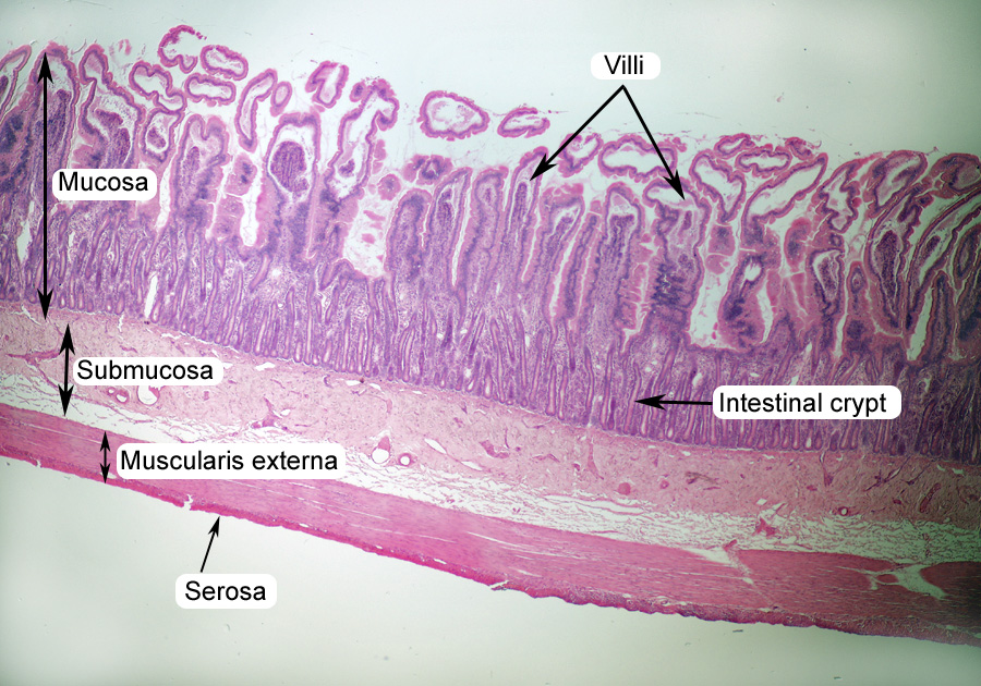
› file › 76838716The Eukaryotic Cell Cycle and Cancer In Depth ... - Course Hero Using the cell cycle diagram on the right and both links in the center purple circle, complete the table below for each phase. Use bullet points and focus on major events that occur during each phase, checkpoint, and regulatory process. Complete the entire row before moving on to the next phase. Digestive: The Histology Guide This is a diagram which shows the villi of the small intestine, as indicated by the arrows in the diagram above, at higher magnification. The main functions of the small intestine are digestion, absorption of food and production of gastrointestinal hormones. The small intestine is 4-6 metres long in humans.
Villi - SmartDraw Villi. Create healthcare diagrams like this example called Villi in minutes with SmartDraw. SmartDraw includes 1000s of professional healthcare and anatomy chart templates that you can modify and make your own. 70/71 EXAMPLES. EDIT THIS EXAMPLE.

Diagram of villi
What Is Chorionic Villi? - Sampling & Definition - Video ... Chorionic villi is a membrane full of blood vessels that surrounds the embryo. This membrane allows the exchange of nutrients and oxygen and waste and carbon dioxide between the mother and the ... Absorption - Digestive system - GCSE Biology (Single ... The villi. The villi (one is called a villus) are tiny, finger-shaped structures that increase the surface area. They have several important features: PDF Cambridge International Examinations Cambridge ... 6 The diagram shows a cell. Which structure is not present? A cell membrane B cell wall C cytoplasm D nucleus 7 The diagram shows blood passing through an arteriole into a capillary. Part of the capillary wall has been cut away to show the blood. Q P direction of blood flow What is the level of organisation of the structures labelled P and Q? P Q
Diagram of villi. quizlet.com › 18983293 › digestive-system-flash-cardsDigestive System Flashcards - Quizlet villi. tiny, finger-like structures that protrude from the wall of the intestine. pancreas. ... Diagram and label the internal structures of the eye, and give the ... en.wikipedia.org › wiki › AmnionAmnion - Wikipedia The amnion is a membrane that closely covers the human and various other embryos when first formed. It fills with amniotic fluid, which causes the amnion to expand and become the amniotic sac that provides a protective environment for the developing embryo. Villi - Small Intestine - SmartDraw Villi - Small Intestine. Create healthcare diagrams like this example called Villi - Small Intestine in minutes with SmartDraw. SmartDraw includes 1000s of professional healthcare and anatomy chart templates that you can modify and make your own. 69/71 EXAMPLES. EDIT THIS EXAMPLE. CLICK TO EDIT THIS EXAMPLE. Microvilli- Definition, Structure, Functions and Diagram Figure: Diagram of Microvilli. Microvilli form a rather polymorphic class of surface protuberances that are regularly packed in some tissues and loosely positioned in others. Generally, they are shorter and smaller in diameter than cilia. They are commonly about 0.1 µm diameter and range in length from a fraction of a micrometer to about 2 µm.
Villi in the Small Intestine - Biology | Socratic The process that the nutrients move into the villi is diffusion. Source, TommyIX, 2013 The picture above is a diagram of what is inside the villus. It explains what kind of nutrients is absorbed by the blood capillary which is glucose, amino acids (and can also be nucleotides) and by the lacteal which is fatty acids and glycerol. Diagram Of Villi In Small Intestine - DiagramSketch Diagram of villi in small intestine. The goblet cells contain many microvilli. The small intestine is part of the digestive system and is vital for breaking down and absorbing nutrients. The small intestine is not flat or. The large surface area helps in the rapid absorption of digested food. Together with the esophagus large intestine and the ... What is the Difference Between Villi and Microvilli ... Villi are small, finger-like projections extending into the lumen of the small intestine. In humans, the villus is approximately 0.5-1.6 mm in length. Each villus contains many microvilli, projecting from the epithelial cells of the intestine. Moreover, villi and microvilli collectively form the brush border. Exploring the villus - PubMed Central (PMC) This diagram represents the types of bone-marrow cell derivatives operative within the lamina propria. They include (in cerise) the subepithelial myofibroblast system (MYF); pericytes (green) supporting the subepithelial capillaries and main vasculature of the villi (artery, red: vein, blue); the lacteal (L) supported by smooth muscle cells (SM) and (purple) the muscularis mucosae (MM).
› articles › 320014Digestion: Anatomy, physiology, and chemistry Jan 11, 2018 · Share on Pinterest A diagram of the human digestive system. ... Tiny finger-like projections called villi stick out from the walls of the duodenum and increase its surface area. Villi maximize the ... A Labelled Diagram Of Digestive System with Detailed ... The diagram below shows the structure and functions of the human digestive system. Let learn the different parts of the human digestive system. Mouth — It includes teeth, salivary glands and tongue. It is the beginning of the digestive tract and the process of digestion begins from the mouth, where teeth help by breaking and grinding the food ... villus | anatomy | Britannica villus, plural villi, in anatomy any of the small, slender, vascular projections that increase the surface area of a membrane. Important villous membranes include the placenta and the mucous-membrane coating of the small intestine. The villi of the small intestine project into the intestinal cavity, greatly increasing the surface area for food absorption and adding digestive secretions. Small Intestine: Anatomy, Function, and Treatment Intestinal villi: The mucous folds in the small intestine are lined with multitudes of tiny finger-like projections that protrude into the opening of the small intestine. These villi are covered with absorptive epithelial cells that take up nutrients from the lumen and transport nutrients into the blood.
Transverse Section of Villi of Small Intestine | ClipArt ETC
Draw the diagram of villi in small intestine and label its ... Identify the cells whose secretion protects the lining of gastro-intestinal tract from various enzymes. Draw the diagram of villi in small intestine and label its parts.
Small Intestine Color Images
How to draw structure of Villi step by step for ... - YouTube How to draw structure of Villi step by step for class Student in easy way. Any student can draw this villi Diagram easily.😇 𝕊𝕦𝕓𝕤𝕔𝕣𝕚𝕓𝕖 :- ...

Chapter 22. Week 3 of Development: Trophoblast and Villus ...
Villus - Definition, Structure, Role, and FAQs Villi Diagram. Below given picture is the villi diagram: [Image will be uploaded soon] Role of Villi. The role of villi or villi function are stated below: The villi along with the microvilli support in increasing the intestinal adsorbent surface area by approximately 30-fold and 600-fold, respectively.

BGDA Practical - Placenta and Fetal Membranes - Embryology
Endoscopic diagnosis of celiac disease - Endoscopy Campus Figure 6: Upper left image presenting the initial diagnosis of celiac disease with fissures on the folds and absence of villi when the mucosa was visualized using NBI and near focus in the right image. The lower images were obtained from the same patient after five months of gluten free diet. The lower left image presents regular folds and the right image using NBI and near focus small, but ...
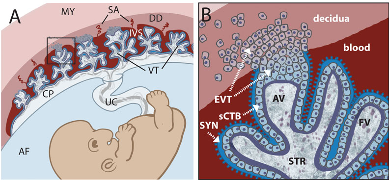
Trophoblast - Embryology
Villi diagram - Healthiack Villi diagram This summary article is displaying Villi diagram … Please click on the diagram(s) to view larger version. You are welcome to browse our website for more details on this specific topic. Best viewed on 1280 x 768 px resolution in any modern browser. This article is about Villi diagram … All pictures are […]

Intestinal villi, light micrograph - Stock Image - C022 ...
villi structure diagram - gkcwpc.org Label the following on a diagram of a villi: capillary, epithelial cell, lacteal, and goblet cell. 2. Since the villi arise from trophoblast cells, their chromosome structure is the same as the fetus's.

Biology - The Digestive Sytem: Draw and label a diagram of ...
The human digestive system - Animal ... - BBC Bitesize The surface of the small intestine wall is folded, and has projections called villi. Villi is the plural of villus. The epithelial cells that cover each villus themselves have projections called...
Digestive
Villi: Function, Definition & Structure - Video & Lesson ... The next feature used to increase surface area is called a villus (plural: villi ). The villi are small, finger-like projections about a millimeter in length that protrude from the circular folds....
Lesson 13 diffusion
What are villi? What is their location and function ... The tiny projections on the inner surface of the small intestine which help in absorbing the digested food are called villi. These helps to increase the surface area of intestinal walls. Location. These are located in the inner walls of the small intestine. Functions


Comments
Post a Comment