40 nerves of the foot and ankle diagram
Foot & Ankle Tendons: Anatomy, Function & Injuries Damage to the foot and ankle tendons are a common cause of foot pain, typically caused by overuse, overstretching or an injury. Tendons are thick bands of tissue that connect muscles to bone. When a muscle contracts, the tendon pulls on the bone causing the joint to move. There are a number of tendons located in the foot and ankle all ... Foot Condition By Area | Top of Foot | Foot Pain Diagram ... Match the corresponding numbers on the foot diagram below for a list of conditions that may be causing your foot and ankle pain. This is meant for educational purposes only. If you're having a problem with your foot or ankle, visit a podiatrist - a foot and ankle specialist! Top (Dorsal) View of Foot & Ankle Number 1 and 2:
Normal Anatomy and Compression Areas of Nerves of the Foot ... The anatomy of the nerves of the foot and ankle is complex, and familiarity with the normal anatomy and course of these nerves as well as common anatomic variants is essential for correct identification at imaging. Ultrasonography (US) and magnetic resonance (MR) imaging allow visualization of these nerves and may facilitate diagnosis of ...

Nerves of the foot and ankle diagram
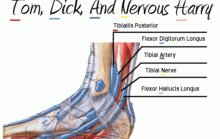
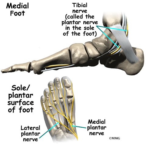
orthopaedia.com › page › Anatomy-of-the-Foot-AnkleAnatomy of the Foot and Ankle - OrthoPaedia Nerves of the Foot. There are five main nerves that run past the ankle into the foot (Figure 17). All five of these are derived from two nerves that originate from the lumbar spine. The sciatic nerve branches into four of the five primary nerves of the foot. Two segments of the sciatic nerve branch before the knee joint: the tibial nerve and peroneal nerve. PDF Basic Structure and Function Ankle and Foot There are six nerves associated with the motor and sensory functions of the foot and ankle. They are: Nerve Innervations: Superficial peroneal nerve (L4-S1) Lateral plantar nerve (S2-S3) Medial plantar nerve (L5-S3) Blood Supply to the Foot - Foot & Ankle - Orthobullets provides blood supply to plantar foot and toes. branches. plantar digital arteries. plantar metatarsal arteries. Arcuate artery. is a vascular arch that runs in the dorsal midfoot deep to the extensor tendons. gives off dorsal metatarsal arteries that run in the 2nd, 3rd and 4th intermetatarsal spaces.
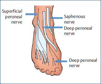
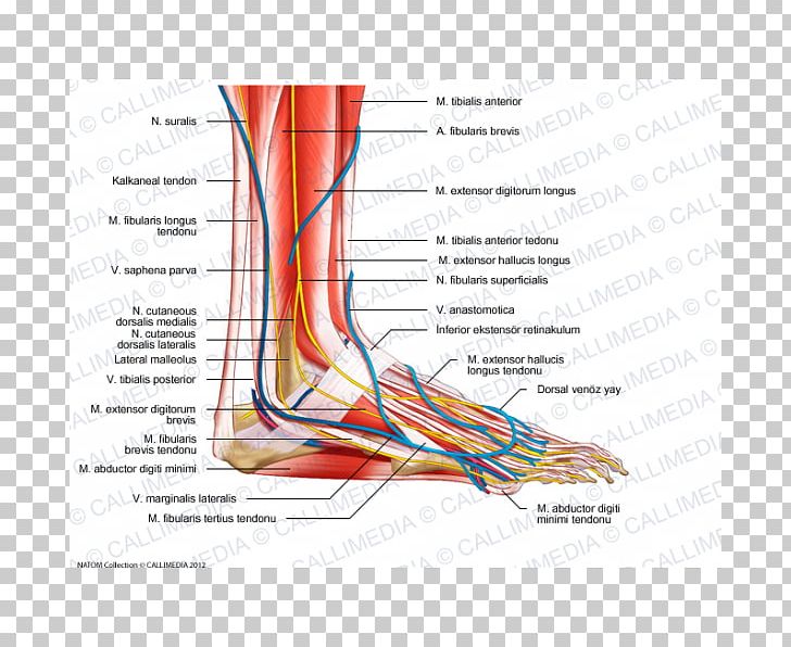
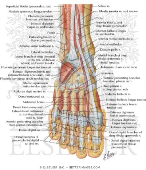
Nerves of the foot and ankle diagram. Nerves Of The Leg & Foot - Everything You Need To Know ... Dr. Ebraheim's educational animated video describes the nerves of the lower leg in a very easy and simple animation.Lateral cutaneous nerve of the calfSural ... Nerves of Foot - Earth's Lab The deep fibular nerve is parallel and lateral to the tendon of the extensor hallucis longus muscle and goes inside the dorsal aspect of the foot on the lateral aspect of the dorsalis pedis artery. The nerve produces a lateral branch just distal towards the ankle joint, which stimulates the extensor digitorum brevis from its deep surface. Ankle and foot anatomy: Bones, joints, muscles | Kenhub Ankle anatomy. The ankle joint, also known as the talocrural joint, allows dorsiflexion and plantar flexion of the foot. It is made up of three joints: upper ankle joint (tibiotarsal), talocalcaneonavicular, and subtalar joints. The last two together are called the lower ankle joint. › anatomy-foot-ankleFoot Ankle Anatomy, Pictures, Function, Treatment, Sprain Pain Oct 17, 2010 · The arteries supplying the ankle joint are branches of the anterior and posterior tibial arteries as well as the peroneal artery. The nerves of the ankle are derived from the deep and superficial peroneal nerves, the tibial nerves, and the sural and saphenous nerves. The Foot Bones of the foot as seen from the medial (arch) side.
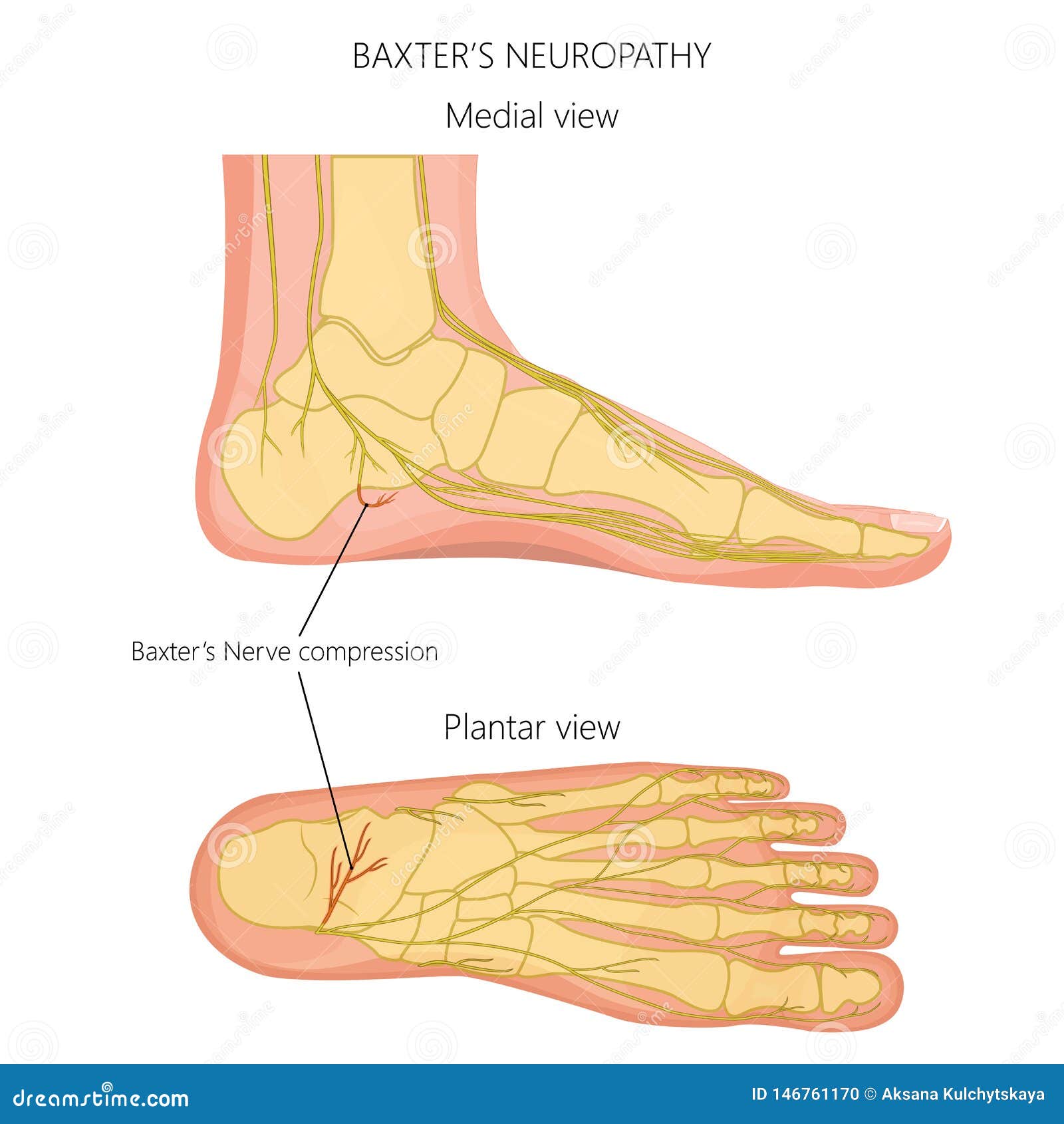
Neuritis and Neuromas of the Foot and Ankle - FootEducation Neuritis and Neuromas of the Foot and Ankle. Edited by Eric Malicky MD . Clinical Presentation. Injury or irritation to nerves in the foot and ankle often creates pain and/or numbness. They occur as a result of an injury to a specific nerve. Nerve damage can result from the original injury, casting/wrap, or surgery. Are Nerve Problems Causing Your Foot Pain? Four common nerve problems can cause foot pain: Morton's neuroma, tarsal tunnel syndrome, diabetic peripheral neuropathy, and a pinched nerve. You'll probably know when trouble strikes. Nerve problems often trigger burning or shooting pain. And the sensation can be so intense that it can rouse you from a deep sleep. A Complete Guide To The Nerves In Your Feet - Foot Vitals Problems with nerves in the feet are very common. Many times, an injured nerve will cause intense pain and heat felt within the foot. Nerves act as a network, communicating important information from the foot to the brain. Learn more about the various conditions and problems that can affect the nerves in the foot. Lower limb anatomy: Bones, muscles, nerves, vessels | Kenhub Ankle and foot. Last but not least, let's tackle the ankle and foot anatomy. The ankle (talocrual) joint is a hinged joint capable of plantarflexion and dorsiflexion. It is composed of three bones: tibia, fibula, and talus (ankle bone). Out of the three, the fibula only plays a secondary and functional role, facilitating the movement of the ...
› anatomy › nervousNerves of the Leg and Foot | Interactive Anatomy Guide Jul 03, 2018 · All of these nerves extend as branches of nerves in the leg that pass through the ankle and into the foot. The sural nerve branches from the tibial and common fibular nerves and is responsible for feeling on the outside of the foot and the small toe. The medial and lateral plantar nerves are the two largest nerves in the bottom of the foot. Normal Anatomy and Compression Areas of Nerves of the Foot ... various nerves about the ankle and foot. It is also important to recognize common anatomic vari-ants. In this article, we review the normal anat-TEACHING POINTS During ankle dorsiflexion, excessive traction may occur along the exit point of the superficial peroneal nerve, injuring it and causing secondary neuroma formation at this level. The nerve Lower limb arteries and nerves: Anatomy, branches | Kenhub Ankle and foot. The ankle region is innervated by articular branches of the tibial and deep fibular nerves. Regarding the nerves of the foot, we have the following: dorsal digital nerves, proper plantar digital nerves, lateral dorsal cutaneous nerve, and plantar (medial and lateral) nerves. › foot-and-ankle › 7004Nerves of the Foot - Foot & Ankle - Orthobullets 3%. (71/2759) 3. It is the terminal branch of the superficial peroneal nerve; injury leads to reduced sensation over medial aspect of great toe. 83%. (2284/2759) 4. It is the terminal branch of the deep peroneal nerve; injury leads to first interphylangeal joint flexion weakness. 3%.
Ankle Block - Landmarks and Nerve Stimulator Technique ...
Foot Pain Diagram - Why Does My Foot Hurt? Foot Pain Diagram. Written By: Chloe Wilson BSc(Hons) Physiotherapy Reviewed By: FPE Medical Review Board A foot pain diagram is a great tool to help you work out what is causing your ankle and foot pain. There are a whole range of structures e.g. bones, muscles, tendons and nerves which will each give slightly different foot pain symptoms.

Foot & Ankle Nerve Injuries | Orthopaedic Associates of St ...
iytmed.com › nerves-in-foot-and-ankleNerves in Foot and Ankle - IYTmed.com The nerves of the foot assistance move the body and keep balance both while it’s moving and at rest. All these nerves extend as branches of nerves in the leg that travel through the ankle and into the foot. The sural nerve branches from the tibial and common fibular nerves and is responsible for feeling on the outside of the foot and the little toe. The median and lateral plantar nerves are the 2 biggest nerves in the bottom of the foot.

13 Ankle anatomy ideas | ankle anatomy, anatomy, muscle anatomy
Blood Supply to the Foot - Foot & Ankle - Orthobullets provides blood supply to plantar foot and toes. branches. plantar digital arteries. plantar metatarsal arteries. Arcuate artery. is a vascular arch that runs in the dorsal midfoot deep to the extensor tendons. gives off dorsal metatarsal arteries that run in the 2nd, 3rd and 4th intermetatarsal spaces.
The Foot – Advanced Anatomy 2nd. Ed.
PDF Basic Structure and Function Ankle and Foot There are six nerves associated with the motor and sensory functions of the foot and ankle. They are: Nerve Innervations: Superficial peroneal nerve (L4-S1) Lateral plantar nerve (S2-S3) Medial plantar nerve (L5-S3)
Nerves of the foot-02 stock vector. Illustration of mortons ...
orthopaedia.com › page › Anatomy-of-the-Foot-AnkleAnatomy of the Foot and Ankle - OrthoPaedia Nerves of the Foot. There are five main nerves that run past the ankle into the foot (Figure 17). All five of these are derived from two nerves that originate from the lumbar spine. The sciatic nerve branches into four of the five primary nerves of the foot. Two segments of the sciatic nerve branch before the knee joint: the tibial nerve and peroneal nerve.

Nerves of the Foot - Foot & Ankle - Orthobullets

Compression Neuropathy - Lincoln Park | Lakeview Chicago, IL ...

The saphenous nerve in foot and ankle surgery: Its variable ...

a Anatomical dissection of the tibial nerve and its branches ...
In the Foot and Ankle Operation Theater | Musculoskeletal Key

Tarsal tunnel - Wikipedia
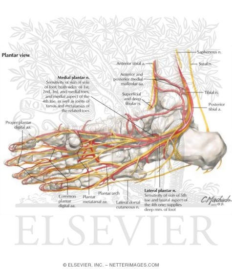
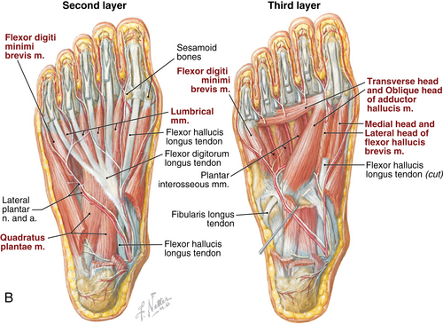
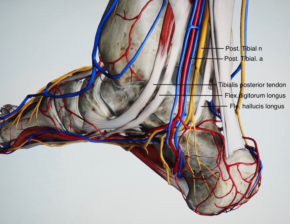
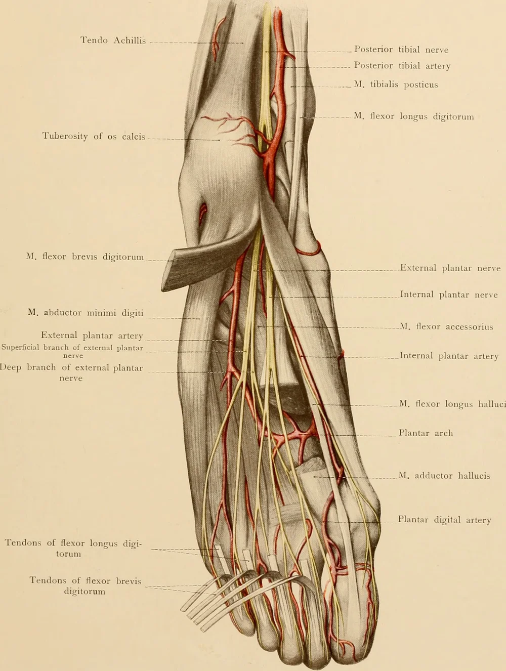
Ankle and Foot: Arteries and Nerves of the Sole

Foot Anatomy - Podiatrist San Angelo, TX

Normal Anatomy and Compression Areas of Nerves of the Foot ...

The Tibial Nerve - Course - Motor - Sensory - TeachMeAnatomy
Foot and ankle | Musculoskeletal Key

Foot and Ankle Pain Treatment in Schertz | BioMotion PT

The leg, ankle, and foot - Knowledge @ AMBOSS

Foot and Ankle | Musculoskeletal Key

Foot and ankle issues | Northern Arizona Healthcare

Anatomy and Injuries of the Foot and Ankle Anatomical Chart ...
BATS - Better Anaesthesia Through Sonography

Anatomy of Ankle and Foot. Overview Bones of Ankle and Foot ...

Anatomy of the Foot and Ankle | OrthoPaedia
Foot Nerve Human Anatomy Muscle PNG, Clipart, Anatomy, Angle ...
Nerve Blocks of the Foot and Ankle — Downeast Emergency Medicine

Ankle Block - Landmarks and Nerve Stimulator Technique ...

Anatomical dissection of the cutaneous nerves of the foot and ...

Nerves and arteries of the foot (preview) - Human Anatomy | Kenhub

5 Anatomy of the distal tibial nerve and its branches (Image ...

Muscles, Arteries, and Nerves of Front of Ankle and Dorsum of ...

Anatomy of the Foot and Ankle | OrthoPaedia

Ankle Anterolateral Approach - Approaches - Orthobullets
Foot Anatomy - eOrthopod.com

Normal Anatomy and Compression Areas of Nerves of the Foot ...

Normal Anatomy and Compression Areas of Nerves of the Foot ...

Stock Ankle: Normal Anatomy — Illustrated Verdict
Muscles, Arteries, and Nerves of Front of Ankle and Dorsum of ...

Low Ankle Sprains - Yakima Foot & Ankle

How to Keep Walking With MS Foot Drop | Everyday Health


Comments
Post a Comment