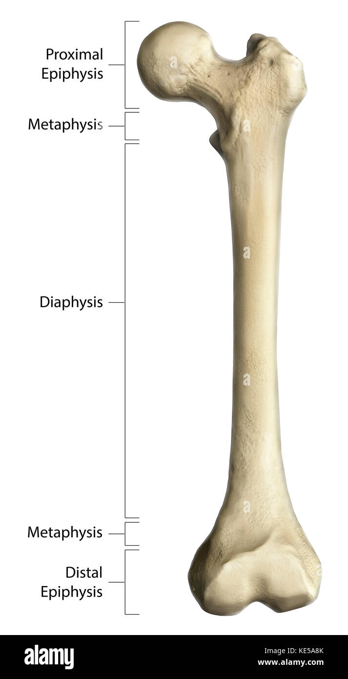
43 where in the diagram is the distal epiphysis
definition of phalanges by The Free ... - The Free Dictionary (2) In distal and middle phalangeal lesions, the conventional procedure is transverse amputation, including the head of the proximal neighboring joint of the diseased phalanx, and closing the defect with dorsal and ventral flaps. The Anatomy of Bones - Quiz 2 - Free Anatomy Quiz In the diagram the 'metaphysis' is indicated by label number : A long, tunnel-like passage is called a : What do we call a large, rounded articular process? Which label in the diagram indicates the\'medullary cavity' : An 'eminence' is : The term 'epicondyle' refers to : In the diagram the 'distal epiphysis' is indicated by label number :
Epiphyseal cortical irregularity | Radiology Case ... Scroll Stack. Frontal. There is mild irregularity with fragmentation of the medial aspect of the distal femoral epiphysis, best appreciated on the AP projection. From the case: Epiphyseal cortical irregularity. X-ray.
Where in the diagram is the distal epiphysis
Salter–Harris fracture - Wikipedia A Salter–Harris fracture is a fracture that involves the epiphyseal plate or growth plate of a bone, specifically the zone of provisional calcification. It is thus a form of child bone fracture.It is a common injury found in children, occurring in 15% of childhood long bone fractures. This type of fracture and its classification system is named for Robert B. Salter and William H. Harris who ... Epiphysis - Explanation, Types, Bones, Similarities and ... The distal epiphysis is also covered in articular cartilage, allowing bones to move freely at the joints without rubbing against one another. The functionality of Epiphysis and distal epiphysis is distinguishable; the traveling of the bones to the joints is made easy with the help of the same. It can get easily affected and get torn. Anatomy And Physiology Questions - ProProfs Anatomy And Physiology Questions - The Skeletal System: Bone Tissue. Everything about bones in cellular level. Linked to the 'Childhood' case unit. 1. This is a structure of a long bone that stores energy. 2. This is the region of a long bone that articulates with other bones. 3. This is the shaft of a logn bone.
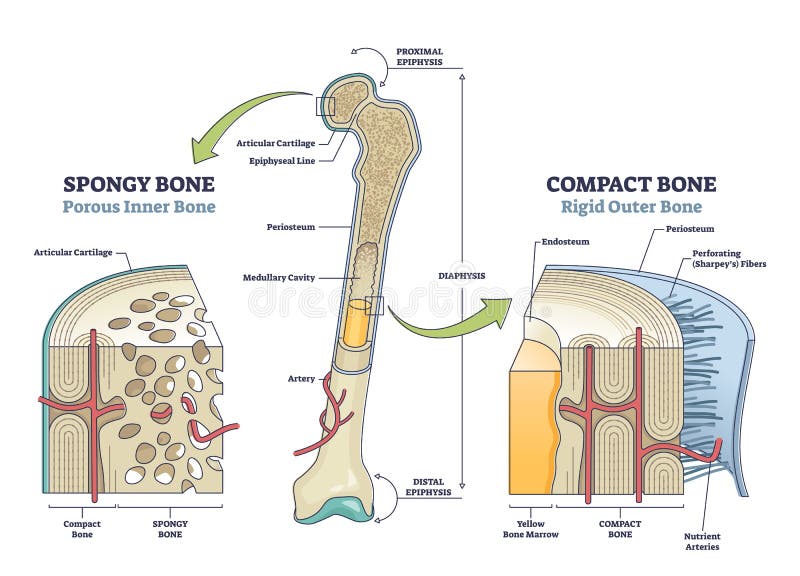
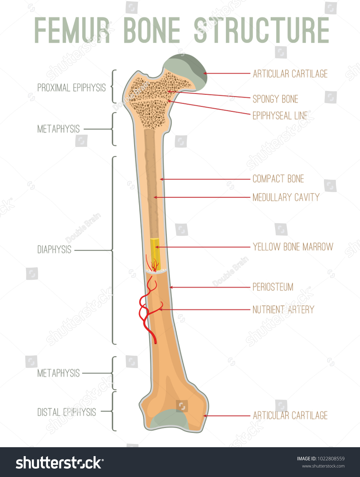
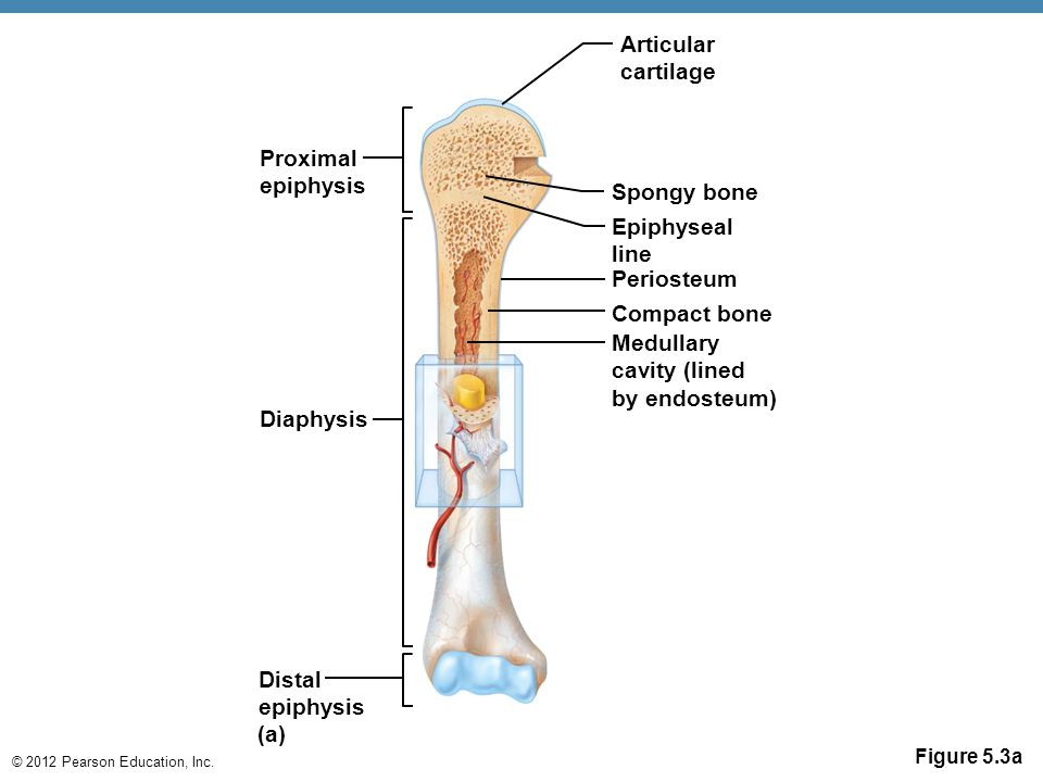
Where in the diagram is the distal epiphysis. 3D model with anatomical sections: Distal epiphysis ... Download scientific diagram | 3D model with anatomical sections: Distal epiphysis, distal metaphysis, shaft, trochanter region and femur head. from publication: The effect of CT slice spacing on ... CLINICAL EXAMINATION AND PHYSICAL ASSESSMENT OF HIP … Pediatric and adolescent pathologies, such as Legg‐Calve‐Perthes (typical age of onset is 3 to 12 years old) and Slipped Capital Femoral Epiphysis (average age of 12.1 years for girls and 14.4 years for boys) will significantly differ in athlete age compared to acetabular labral tear (ALT) (adolescents to older adults) and hip ... Secondary epiphyses of the mammalian pisiform and ... Download scientific diagram | Secondary epiphyses of the mammalian pisiform and calcaneus. (A) Ulnar view of a 1-month-old mouse forepaw generated from micro-CT. Note the elongated pisiform (white ... Long Bone Anatomy: Structure and Parts of Long Bones The labels include proximal epiphysis, proximal metaphysis, diaphysis (bone shaft), distal metaphysis, distal epiphysis, and epiphyseal line (x2). Structure of an adult human long bone The following image gets into a little more detail in regard to human long bone structure.
36 Where in the diagram is the distal epiphysis a A b B c ... Ans: The diagram is depicting intramembraneous ossification in a child's skull. In number 1 there is the development of an ossification center. In picture 2 calcification begins. In picture 3 trabeculae begins to form. In picture 4 the periosteum develops Difficulty: medium. dyjf.docx - Where in the diagram is the distal epiphysis ... View dyjf.docx from SP 21 at Technical University of Mombasa. Where in the diagram is the distal epiphysis? D In which region of the diagram would you find the medullary cavity? C Where in the Complete the following: Label the diagram to the right: a ... Complete the following: Label the diagram to the right: a. diaphysis b. proximal and distal epiphysis c. periosteum d. spongy bone e. compact bone f. medullary cavity Solved EXERCISE 5.3 Anatomy of a Long Bone As ... - Chegg Transcribed image text: EXERCISE 5.3 Anatomy of a Long Bone As opposed to the other categories of bone, all long bones share several characteristics 1 Label the diagram with the following terms: Articular cartilage (distal) Articular cartilage (proximal) Compact bone Duphysis Distal epiphysis Endosteum Epiphyseal line (distal) Epiphyseal lines (proximal) Medullary Cavity Periosteum Proximal ...
What Is the Difference between the Epiphysis and Diaphysis? Rebecca Harkin Date: February 23, 2022 The anatomy of a bone.. The epiphysis and diaphysis are different parts of a long bone, or a bone found in a limb. Knobby ends of a long bone are referred to as the epiphyses, and the diaphysis is the shaft or middle section of the long bone. Complete the following: Label the diagram: a. diaphysis ... Complete the following: Label the diagram: a. diaphysis b. proximal and distal epiphysis c. periosteum d. spongy bone e. compact bone f. medullary cavity A&P Homework 1 Study Flashcards - Quizlet Where in the diagram is the distal epiphysis? a. A b. B c. C d. D e. E. d. 20. Where in the diagram can you find the medullary cavity? a. A b. B c. C d. D e. E. c. 21.Where in the diagram can you find red bone marrow in an adult? a. A and B b. B and D c. A and D d. C e. E. c. 22. Where in the diagram is the metaphysis? Difference Between Epiphysis and Diaphysis | Compare the ... The gross structure of the long bone consists of many parts; proximal and distal epiphysis, the spongy bone and the diaphysis consisting of the medullary cavity, endosteum, periosteum and the nutrient foramen. Thus, the anatomical structure of the long bone is divided into two main parts. They are the epiphysis and the diaphysis.
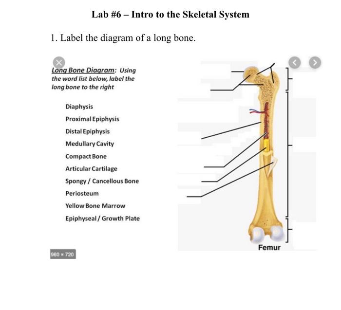
Solved Lab #6 - Intro to the Skeletal System 1. Label the ...
Study 70 Terms | Biology Flashcards - Quizlet Where in the diagram is the distal epiphysis? D. In which region of the diagram would you find the medullary cavity? C. Where in the diagram can you find red bone marrow in an adult? A & B. Where in the diagram is the metaphysis? B. What structure in the diagram is the only place on a long bone NOT covered by the periosteum? E.

Epiphysis Diagram | Quizlet
Distal Epiphysis: study guides and answers on Quizlet Periosteum 7. Promixal Epiphysis 8. Diaphysis 9. Distal Epiphysis. Explain the advantage for melanin granules being located in the deep layer of the epidermis. Melanin is used to protect the cell's nucleus from UV radiation. Figure 12.2 features associated with the microscopic structure of bone. 1. Spongy Bone 2.

A8-22, right distal epiphysis of humerus in cranial (1a) and ...
Comments on Fractures of the Radius - Prep4usmle.com -Colles fracture occurs when a person falls on the outstretched hand. It's a distal radial fracture. YOu will see it is deformed adn painful. The main lesion is a dorsally displaced, dorsally angulated fracture of the distal radius.-Smith fracture is also called a "reverse colles fracture".Its a fracture of the distal radial metaphysis.

Diagram Of Human Bone Anatomy-vektorgrafik och fler bilder på ...
Femur bone anatomy: Proximal, distal and shaft | Kenhub 2022-02-17 · Varus inclination of the proximal tibia or the distal femur does not influence high tibial osteotomy outcome. Knee Surgery, Sports Traumatology, Arthroscopy, 17(4), 390-395. doi: 10.1007/s00167-008-0708-6; Melbourne, T. Clinical Practice Guidelines: Slipped upper femoral epiphysis (SUFE) - Emergency Department. Retrieved from ...

Visual Study Guide Part 1 - Last Quiz of Bone Unit Diagram ...
The Hidden Threat: The Stubbed Toe and Finger - Cincinnati ... The physis, or growth plate, is the cartilage allowing growth of the bone, which shows up on x-rays as a darker zone between the epiphysis at the end of the bone and the metaphysis, which is the shaft portion on the other side of the physis (see diagram 1). A distal phalanx is the outmost bone of the bones in each finger or toe.

Raman spectra of articular surface of femoral bones of rabbit ...
epiphysis | Definition, Anatomy, & Function | Britannica epiphysis, expanded end of the long bones in animals, which ossifies separately from the bone shaft but becomes fixed to the shaft when full growth is attained. The epiphysis is made of spongy cancellous bone covered by a thin layer of compact bone. It is connected to the bone shaft by the epiphyseal cartilage, or growth plate, which aids in the growth of bone length and is eventually replaced ...

Left: distal epiphysis of the right femur in axial view ...
Anatomy Ch.6 Bone Tissue Flashcards | Quizlet In the diagram, where is the zone of resting cartilage and does it demonstrate a proximal epiphysis or a distal epiphysis? D; proximal epiphysis Which of the following selections correctly lists the sequence of events that occur during intramembranous ossification?

Cranial view of the distal epiphysis of the left humerus of ...
Human A&P Ch. 6 Flashcards - Quizlet B) epiphysis C) metaphysis D) periosteum E) marrow 3. This is the shaft of a long bone. A) diaphysis B) epiphysis C) metaphysis D) periosteum E) marrow Ans: A Section Reference 1: 6.2 Structure of Bone 4. This is a layer of hyaline cartilage that reduces friction between bones involved in a joint. A) periosteum B) distal epiphysis C) nutrient ...
Cunningham's Text-book of anatomy. Anatomy. 254 OSTEOLOGY ...
Nodes of Ranvier: Function and Definition - Video & Lesson ... 2021-11-01 · Nodes of Ranvier are vital breaks in a neuron's myelin coating. Explore the parts of a neuron, the definition of these nodes, and discover their primary functions and structure.

Osteon Fotografier, bilder och bildbanksfoton - iStock
Epiphysis - an overview | ScienceDirect Topics Approximately 6% to 15% of long bone fractures occurring in children younger than 16 years of age involve the epiphysis. 185,207,227 Because the epiphysis is responsible for longitudinal bone growth, injury disrupting vascular supply to the epiphysis or metaphysis affects bone growth. The distal tibia, fibula, ulna, and radius are the most ...
Establishment and Characterization of a Clinically Relevant ...
Solved Procedure Part 1 1. Locate these structures and ... Locate these structures and label (the diagram below) the cross section of the long bone: proximal epiphysis, distal epiphysis, epiphyseal plate, articular cartilage, diaphysis, periosteum, compact bone, spongy bone, medullary cavity (9pts) Long Bone 10 . This problem has been solved! See the answer See the answer See the answer done loading.

Osteoporosis and spinal cord injury - Northwest Regional ...
Free Distal Epiphysis Art Prints and Artworks | FreeArt Get Up to 10 Free Distal Epiphysis Art Prints. 9 Distal Epiphysis Art Prints to choose from. New images for 2022, Gallery-Quality Artwork, Free Returns, Ships Same Day.
Skeletal Tissues
A comparison of the semi-pronated distal antebrachial ... Download scientific diagram | A comparison of the semi-pronated distal antebrachial epiphyses of select archosauromorphs and basal archosaurs. A. Archosauromorph, Trilophosaurus buettneri Case ...

Skeletal System Diagram 2 Diagram | Quizlet
Distal Radial Epiphysis Injury - causes, symptoms & treatment The epiphysis is the name given to the rounded end of a long bone also sometimes known as the growth plate. It is the part of the bone which is still growing in young athletes. Injury to the distal radius epiphysis more commonly affects young athletes between the ages of 6 and 10 years old, particularly gymnasts.
An atlas of human anatomy for students and physicians ...
The Human Skeleton - Institute for Emerging Issues The epiphysis is a cartilaginous area of bone growth located near the ends of long bones. As individuals mature, these epiphyses gradually ossify and join the diaphysis, in a timed sequence. In flat bones, such as the skull, growth occurs from the center of the bone. Upon maturation, growth stops and the sutures gradually close. The pubic ...

Drawing illustrates the anatomy at the end of a long bone ...
Endocrine System Development - Embryology 2022-03-09 · Endocrine Pancreas "The transcription factor Pax6 functions in the specification and maintenance of the differentiated cell lineages in the endocrine pancreas. It has two DNA binding domains, the paired domain and the homeodomain, in addition to a C-terminal transactivation domain. The phenotype of Pax6-/- knockout mice suggests non-redundant functions of the …
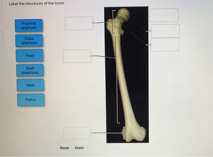
Solved Label the structures of the bone. Proximal epiphysis ...
Sickle cell disease promotes sex-dependent pathological ... 2022-02-25 · (A) Representative microCT image of an entire femoral bone, including a microCT scan of the distal femur with the distal epiphysis outlined in red. (B) Representative three-dimensional heat maps of trabecular morphology in the epiphyseal region of the distal femur in AA, AS, and SS in 3-month-old (left) and 5-month-old (right) male and female mice. A …

LABEL A LONG BONE (HUMERUS) Diagram | Quizlet
The Proximal Tibia1 Epiphysis - tandfonline.com The proximal epiphysis of the tibia is present in some 75 to 80 per cent of all full-term children (ADAIR and ScAhmON, 1921; MENEES and HOLLY, 1932). According to SICK (1902) the proximal tibial epiphysis resembles the distal fe- moral in being rounded initially. To begin with it grows rather rapidly and at

HW #9: Long Bone Anatomy Diagram | Quizlet
What Is Epiphysis of Bone? | Proximal & Distal Epiphysis ... The distal epiphysis is located at the end of the long ... There can be some confusion about the difference between the metaphysis and the epiphysis. The diagram below should help to clarify what ...
Gross anatomy of a long bone, using a femur with annotations ...
Anatomy And Physiology Questions - ProProfs Anatomy And Physiology Questions - The Skeletal System: Bone Tissue. Everything about bones in cellular level. Linked to the 'Childhood' case unit. 1. This is a structure of a long bone that stores energy. 2. This is the region of a long bone that articulates with other bones. 3. This is the shaft of a logn bone.
SKELETAL SYSTEM OVERVIEW OBJECTIVES
Epiphysis - Explanation, Types, Bones, Similarities and ... The distal epiphysis is also covered in articular cartilage, allowing bones to move freely at the joints without rubbing against one another. The functionality of Epiphysis and distal epiphysis is distinguishable; the traveling of the bones to the joints is made easy with the help of the same. It can get easily affected and get torn.

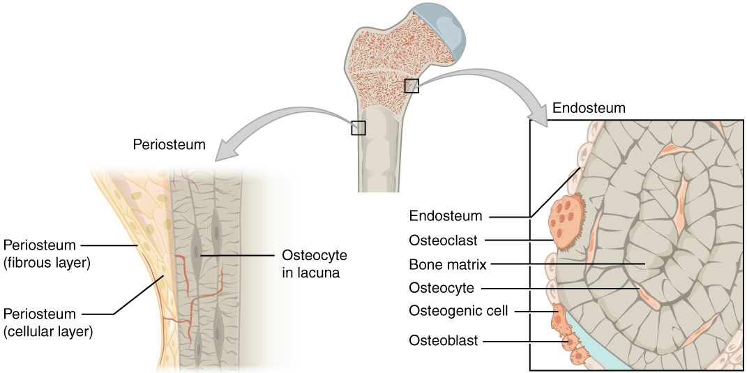
6.3 Bone Structure – Anatomy & Physiology
Salter–Harris fracture - Wikipedia A Salter–Harris fracture is a fracture that involves the epiphyseal plate or growth plate of a bone, specifically the zone of provisional calcification. It is thus a form of child bone fracture.It is a common injury found in children, occurring in 15% of childhood long bone fractures. This type of fracture and its classification system is named for Robert B. Salter and William H. Harris who ...

Proximal Epiphysis Stock Illustrations – 8 Proximal Epiphysis ...

Epiphyseal, proximal epiphysis, diaphysis, distal epiphys...

Individual no. 155: A – in situ, B- skeletal inventory, C ...


What Is Epiphysis of Bone? | Proximal & Distal Epiphysis ...

96 Bone Tissue Illustrations & Clip Art - iStock
Proximal Epiphysis Stock Illustrations – 8 Proximal Epiphysis ...

Anterior view of the distal epiphysis of the right radius ...

Strukturen I En Lång Ben Humerus Röret-vektorgrafik och fler ...

Schematic diagram of system used for measuring streaming ...

Vektor Illustration Systemet Av Ben Tvärsnitt Diagram Med ...

Cranial view of the distal epiphysis of the right humerus of ...

IB DP Language A Language and Literature -sample text ...
Long bone growth and evolution revealed by three-dimensional ...
Vector Illustration Scheme Bone Cross Section Stock Vector ...

Chapter 6 Bone Tissue Diagram | Quizlet
Anatomy skeleton diagrams - ppt video online download

Comparison of the left distal epiphysis of the radius of the ...

Pictures Anatomy & Physiology Midterms Flashcards | Quizlet
Bone Structure – Anatomy and Physiology

Representative 3-D images of the distal epiphysis between 1.5 ...


Comments
Post a Comment