43 compound microscope ray diagram
(i) Draw a neat labelled ray diagram of a compound ... Draw a neat labeled ray diagram of a compound microscope. Explain briefly its working. asked Aug 1, 2019 in Physics by Rk Roy (63.8k points) jee; jee mains; 0 votes. 1 answer (i) Draw a neat labelled ray diagram of an astronomial telescope in normal adjustment . Explain briefly its working . (ii) An astronomical telescope u Types of Microscopes: Definition, Working Principle ... Where, D is the least distinct vision; F is the focal length of the convex lens; Simple Microscope Diagram. Principle of Simple Microscope. The working principle of a simple microscope is that when a sample is placed within the focus of the microscope, a virtual, erect and magnified image is obtained at the least distance of distinct vision from the eye that is held at the lens.
Ray Diagram - Compound Microscope - YouTube How to draw ray diagram of Compound Microscope - Chapter 7 - Lenses - Part 4

Compound microscope ray diagram
eHarcourtSchool.com has been retired Connected Teaching and Learning. Connected Teaching and Learning from HMH brings together on-demand professional development, students' assessment data, and … The compound microscope - how to draw ray diagrams - YouTube An animated presentation showing you how to draw ray diagrams (using simple lens rules) for a compound microscope. This shows how to determine the position a... microscope | Types, Parts, History, Diagram, & Facts ... microscope, instrument that produces enlarged images of small objects, allowing the observer an exceedingly close view of minute structures at a scale convenient for examination and analysis. Although optical microscopes are the subject of this article, an image may also be enlarged by many other wave forms, including acoustic, X-ray, or electron beam, and be received by direct …
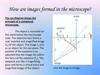
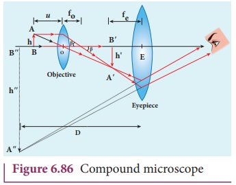
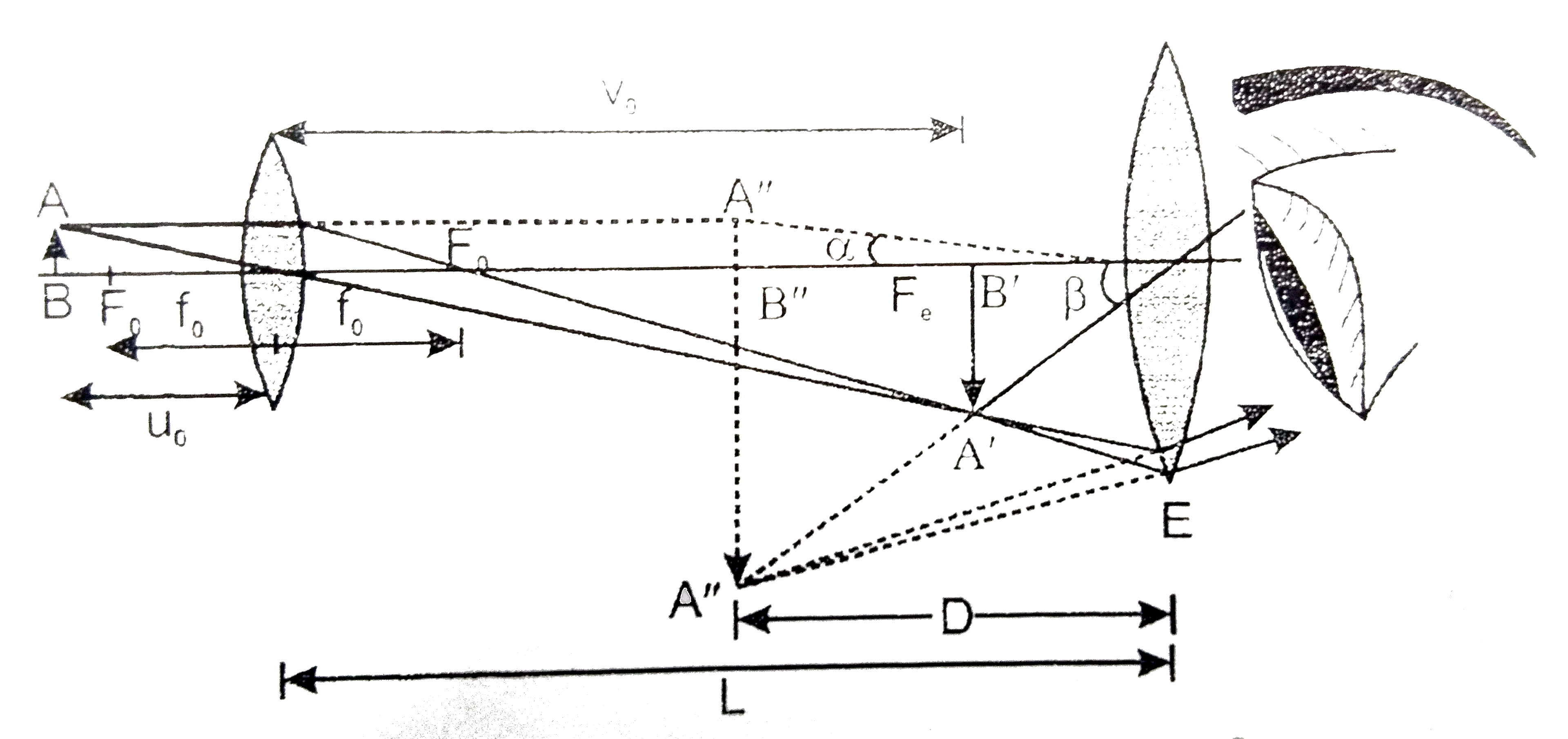
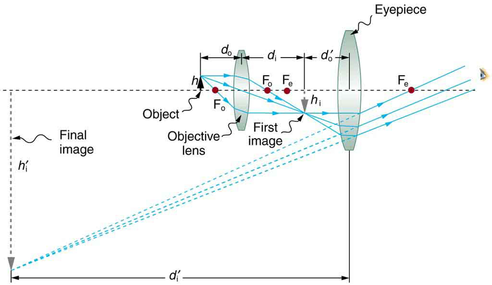
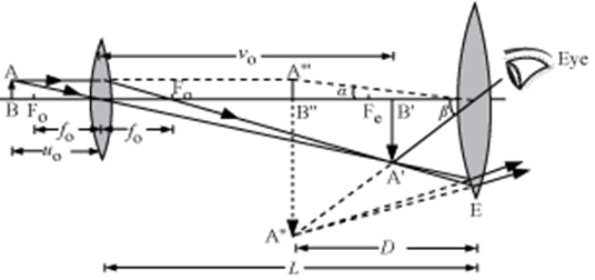
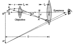
Compound microscope ray diagram. Kingdom Animalia - Different Phylum, Classification ... Kingdom Animalia Different Phylum, Classification, Characteristics Overview. The Kingdom Animalia is a large group that consists of eukaryotic, multicellular organisms that are heterotrophic in nature. As such, they obtain their nutrition from external sources. Although they are unable to produce their own food, which is one of the main defining characteristics of plants, animal cells … Simple Microscope - Definition, Types, Working Principle ... A simple microscope consists of a convex lens of a short focal length. The below figure shows the ray diagram which subsequently forms the image of an object (or we can say a source of light). (Image will be Updated soon) F is the focal length of the lens. An object is placed between the focal length and the centre of the curvature. (a) Draw a labelled ray diagram of a compound microscope ... Best answer (a) Labelled diagram of compound microscope. The objective lens form image A' B' near the first focal point ofeyepiece. (b) Angular magnification of objective lens m0 = linear magnification h'/h where L is the distance between second focal point of the objective and first focal point of eyepiece. What is a Compound microscope? Applications of Compound ... Ray diagram of Compound microscope. The specimen AB is placed just beyond the principal focus Fo' of the objective lens. A ray of light AO from A goes parallel to the principal axis towards the objective lens and converges.
Derive the formula for angular magnification of a compound ... The angular magnification of a compound microscope is the ratio of the angle subtended by the final image at the eye to the angle subtended by the object at the eye, when both are placed at the least distance of distinct vision.This is the required expression for angular magnification. ... Draw the required ray diagram. ... (a) Draw a labelled ray diagram of compound microscope ... (a) Draw a labelled ray diagram of compound microscope, when final image forms at the least distance of distinct vision. (b) Why is its objective of short focal length and of short aperture, compared to its eyepiece? Explain. (c) The focal length of the objective is 4 cm while that of eyepiece is 10 cm. The object is placed at a distance of 6 cm from the objective lens. The Microscope Optical Train - Nikon's MicroscopyU A finite (fixed tube length) microscope optical train is illustrated inFigure 12, which includes the essential optical elements and ray traces defining the relationship between image planes. A specimen located a short distance before the objective front focal plane is imaged through conjugate planes onto the retina of the eye at Image Plane (4) . Draw a Ray Diagram to Show the Working of a Compound ... Draw a ray diagram to show the working of a compound microscope. Deduce an expression for the total magnification when the final image is formed at the near point. In a compound microscope, an object is placed at a distance of 1.5 cm from the objective of focal length 1.25 cm.
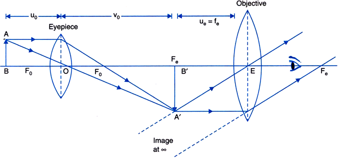
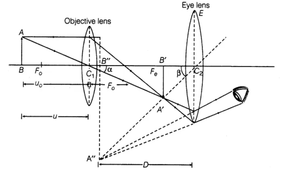
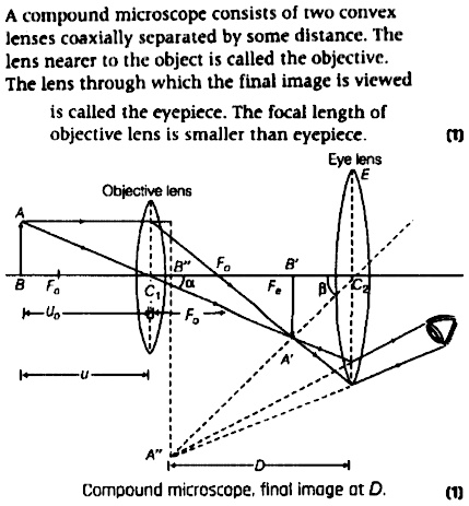
Describe the compound microscope based on the following ... The following ray diagram shows the formation of an image by a compound microscope. The magnifying power of a microscope is the ratio of angle subtended by image to the angle subtended by image placed at least distance of distinct vision. Therefore, from above diagram, m = β α …………….. (1) ∴ α = A B D ……………… (2) Draw a Ray Diagram Showing the Image Formation by a ... Draw a ray diagram showing the image formation by a compound microscope. Hence obtained expression for total magnification when the image is formed at infinity. Advertisement Remove all ads Solution A compound microscope consists of two convex lenses co-axially separated by some distance. The lens nearer to the object is called the objective. Draw a ray diagram of a compound microscope. Write the ... Ray diagram of a compound microscope.When the final image is formed at the least distance of distinct vision,For the image formed at infinity, ue = feand By making focal length of the objective small, the magnifying power can be increased. Compound Microscope: Definition, Diagram, Parts, Uses ... A compound microscope is defined as A microscope with a high resolution and uses two sets of lenses providing a 2-dimensional image of the sample. The term compound refers to the usage of more than one lens in the microscope. Also, the compound microscope is one of the types of optical microscopes.
KOPAL Classes - Compound Microscope Ray Diagram | Facebook
Difference Between Telescope and Microscope The ray diagram for a compound microscope is: Ray Diagram for a compound microscope. Just as it was with telescopes, microscopes are not limited to optical ones either. Electron microscopes use a beam of electrons to look at things at the cellular scale.
Draw a ray diagram showing the image formation by a compound ...
Class 12 Physics Revision Notes for Chapter 9 - Ray Optics ... Consequently, when it is held close to the eye magnified, an erect and virtual image is formed. On the other hand, a compound microscope has two converging lenses, an eyepiece with moderate focal length and large aperture, objective lens of small focal length and short aperture. Telescope. This device is used to observe objects which are far away.
Compound Microscope, Ray Diagram Mistakes. | Physics Forums
Who Invented the Telescope? | Space 26/10/2021 · Lippershey, therefore, gets the credit for the telescope, because of the patent application, while Jansen is credited with inventing the compound microscope. Both appear to have contributed to the ...
Which ray diagram is correct for a Compound microscope ...
optics - Ray diagram of focussing on a compound microscope ... May 19, 2017 · Here is the ray diagram of a compound microscope. So, when we are focussing, we move the objective lens which tweaks the image distance. My doubt is that, shouldn't the image be seen clearly, wheresoever the first real image forms, if within Fe (Focus of the eyepiece lens).

Draw a labelled ray diagram of an image formed by a compound ...
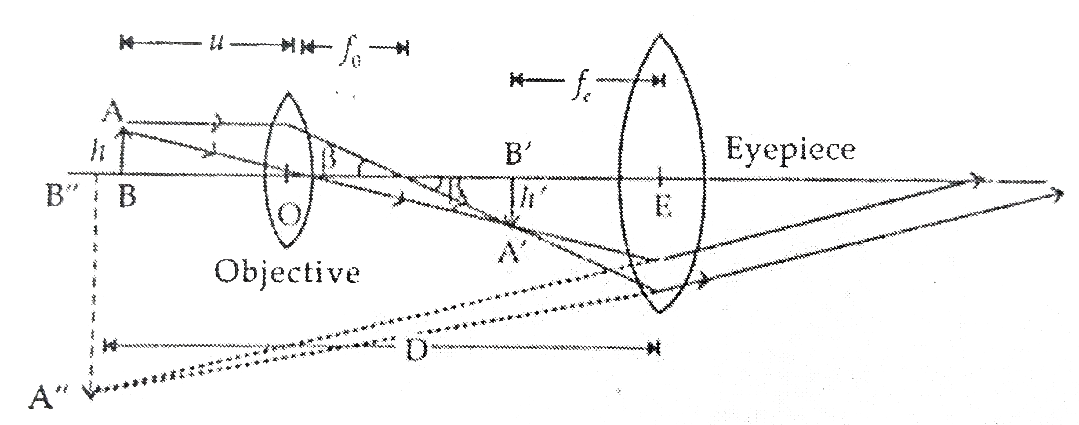
Compound Microscope - Fun Science Working of compound microscope The ray diagram to show the working of compound microscope is shown in figure. A tiny object AB to be magnified is placed in front of the objective lens just beyond its principal focus fo'. In this case, the objective lens O of the compound microscope forms a real, inverted and enlarged image A'B' of the object.

a) Draw a labelled ray diagram of compound microscope, when ...
CBSE NCERT Notes Class 12 Physics Ray Optics Optical ... Simple Microscope. An instrument that gives an enlarged image of a minute object. A simple magnifier or microscope is a converging lens of small focal length. There are 2 types of Microscopes:-. Simple Microscope 2. Compound Microscope. Simple Microscope. The lens is held near the object, one focal length away or less, and the eye is positioned ...
The Compound Microscope - ppt download
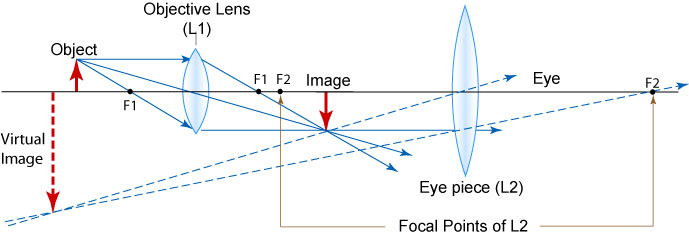
Which ray diagram is correct for a Compound microscope ... Here are two ray diagrams for compound microscope, the first one proposed by the book, and the second one recommended by the teacher: In the first image, the light rays form a real image A'B', which becomes the virtual object for the eyepiece. See, the original rays are carried forward to the eyepiece, which then form a virtual image, A"B".

ICSE Class 8 Physics - Light - Compound Microscope Ray Diagram & Working
Draw a ray diagram of compound microscope when the class 12 ... Draw a ray diagram of compound microscope, when the final image is formed at the minimum distance of distinct vision. Answer Verified 119.7k + views Hint: A compound microscope is an optical instrument used for observing highly magnified images of tiny objects.
Convex lens use - Microscope
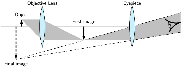
derivation of compound microscope - Physics ... A schematic diagram of a compound microscope is shown in Figure. The lens nearest the object, called the objective, forms a real, inverted, magnified image of the object. This serves as the object for the second lens, the eyepiece,

how to draw Compound Microscope ray diagram step wise how to ...
Compound Microscope | Class 12 Physics Chapter 9 ... Topic compound microscope explains detail working with ray diagram and magnification of compound microscope. Helpful for cbse class 12 physics chapter 9 ray optics. CBSE 12 Physics 01 Electric Charges and Fields 17 Topics 01.01 Electric Charge. 01.02 Conductors, Semiconductors and Insulators.
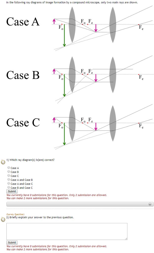
Solved In the following ray diagrams of image formation by a ...
Memristor-based biomimetic compound eye for real-time ... 13/10/2021 · With the hemispherical shaped biomimetic compound eye demonstrated in Fig. 3e, the identical incident angle of 180° along both the x and y …

Ray Diagram of a Compound Microscope | Diagram, Microscope ...
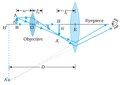
Draw a ray diagram of compound microscope, when final ... Draw a ray diagram of compound microscope, when final image is formed at the minimum distance of distinct vision. Easy Solution Verified by Toppr It consist of two convex lenses, one objective of very small focal length with short aperture. And one Eyepiece with moderate focal length and large aperture.
Which ray diagram is correct for a Compound microscope ...
Compound Microscope- Definition, Labeled Diagram ... The naked eye can now view the specimen at magnification 400 times greater and so microscopic details are revealed. Alternatively, the magnification of the compound microscope is given by: m = D/ fo * L/fe where, D = Least distance of distinct vision (25 cm) L = Length of the microscope tube fo = Focal length of the objective lens
The microscope
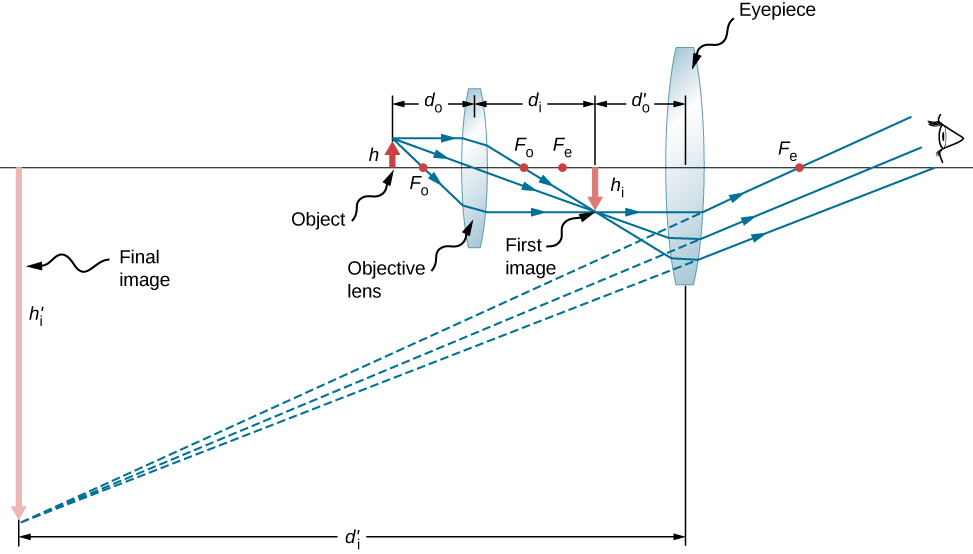
PDF Physics 41: The Compound Microscope distance or image size. Thus, a ray diagram is used to find the final image distance and to determine the total magnification. The overall magnification of a compound microscope is the product of the individual magnifications of each lens: m = mome (1) where the magnification of either lens is given by: m = - q/p (2)

Draw the ray diagram of compound microscope.Tell me the ...
Ray Diagrams for Microscope and Telescope - Wolfram ... Interact on desktop, mobile and cloud with the free Wolfram Player or other Wolfram Language products. Do not show again. Download Wolfram Player. This Demonstration shows the linear magnification for a pair of lenses such as in a microscope or telescope. Contributed by: Volodymyr Holovatsky (Chernivtsi National University, Ukraine) (March 2009 ...
Compound microscope - Optical Instruments
[Term 2] (a) Draw a ray diagram of compound microscope for ... (a) Draw a ray diagram of a compound microscope for the final image formed at least distance of distinct vision? Answer Diagram of Compound Microscope for the final image formed at D: (b) An angular magnification of 30X is desired using an objective of focal length 1.25 cm and an eye piece of focal length 5 cm.

Draw a neat labelled diagram of a compound microscope and ...
Simple microscope - Fun Science Define microscope. Name types of microscope. What is a simple microscope? Give uses of simple microscope. Which type of lens is used as a simple microscope? Explain the principle, construction and working of simple microscope with the help of a ray diagram. Explain magnification of a simple microscope. How it can be increased?
Draw a ray diagram of a compound microscope. Write the ...
microscope | Types, Parts, History, Diagram, & Facts ... microscope, instrument that produces enlarged images of small objects, allowing the observer an exceedingly close view of minute structures at a scale convenient for examination and analysis. Although optical microscopes are the subject of this article, an image may also be enlarged by many other wave forms, including acoustic, X-ray, or electron beam, and be received by direct …
draw a labelled ray diagram showing imagr formation in ...
The compound microscope - how to draw ray diagrams - YouTube An animated presentation showing you how to draw ray diagrams (using simple lens rules) for a compound microscope. This shows how to determine the position a...
Draw a ray diagram to show the working of a compound ...
eHarcourtSchool.com has been retired Connected Teaching and Learning. Connected Teaching and Learning from HMH brings together on-demand professional development, students' assessment data, and …
a) Draw a ray diagram showing the image formation by a ...
Introduction to the microscope A microscope is an example of a ...
Exercises, Telescopes and microscopes, By OpenStax (Page 2/2 ...

Draw a ray diagram to show formation of an image by a ...
Microscopes and Telescopes – University Physics Volume 3
Microscopes | Physics

Optical Instruments: Compound Microscope and its Magnification
Draw the labelled ray diagram for the formation of image by a ...
Microscopes | Physics

Draw a ray diagram for a compoundmicroscope. Derive an ...
Differentiate between compound microscope and astronomical ...

How to Draw Ray Diagram of Compound microscope
a) Draw a ray diagram showing the image formation by a ...
Draw a ray diagram to show the working of a compound ...

Draw the ray diagram of image formation in case of compound ...

optics - Ray diagram of focussing on a compound microscope ...
Draw a ray diagram for the formation ofimage by a compound ...

Draw a labelled ray diagram of an image formed by compound ...

Draw a labelled ray diagram of an image formed by a compound microscope with final image formed ...

Draw a ray diagram for final image formed at distance of ...
MAGNIFICATION IN MICROSCOPE | cell in life

a Draw a ray diagram for the formation of image by a compound ...

Ray Diagram of Compound Microscope (Image at Near Point)


Comments
Post a Comment